
Sympathetic Division of the Autonomic Nervous System
When a threat is presented we have only a few options on how to respond: fight, freeze, or flight. The sympathetic system causes widespread (divergent) effects for many different effector organs spanning various body systems. By connecting various body system through communication it is ensured that the systems are activated together for the common purpose of responding to the threat. More oxygen needs to be inhaled and delivered to skeletal muscle. The respiratory, cardiovascular, and musculoskeletal systems are all activated together. Additionally, sweating cools the body as the water evaporates, releasing the heat that comes from muscle contraction and would otherwise cause the body to overheat. The digestive system shuts down so that blood is not absorbing nutrients when it should be delivering oxygen to skeletal muscles. As you can guess by now the interactions that occur between body systems and the timing of these events are critical for survival in response to the presented threat. To coordinate all these responses, the information about the perceived threat diverges from a limited region of the central nervous system (CNS) to a wide array of ganglia that project to the many effector organs simultaneously. This means that the information that began in a relatively small region of the brain is passed on to a large number of ganglia which in turn pass the information to an even larger array of effector organs.
Think of it like a classroom setting. The teacher represents the start of the signal in the CNS, the preganglionic neuron fiber. She tells one class composed of 20 students about the ANS. This means the information has diverged from 1 to 20 in this one step. The students, representing the postganglionic fibers, each go into a different classroom of 20 students, representing the effector organs, and pass on the information about the ANS. This means the information diverged once more from 20 to 400 in the second step. This really puts into perspective how the signal of the threat is passed very quickly and rather diffusely to initiate body system interaction and regulate the timing of these activities producing a coordinated, systemic change.
The sympathetic division of the autonomic nervous system influences the various organ systems of the body through connections emerging from the thoracic and upper lumbar spinal cord. It is referred to as the thoracolumbar system to reflect this anatomical basis. The preganglionic neuron projects from the lateral grey horn of the thoracic and lumbar regions of the spinal cord to the autonomic ganglion adjacent to the vertebral column via the ventral/motor spinal roots. (Please refer to the diagram given earlier in this module to visualize and refresh your memory of the preganglionic neuron and autonomic ganglion.) The majority of these autonomic ganglia within the sympathetic system belong to a network of sympathetic chain ganglia that runs alongside the vertebral column. The ganglia appear as a series of clusters of neurons linked by axonal bridges – almost like beads on a string. There are typically 23 ganglia in the chain on either side of the spinal column. Because the autonomic ganglion is so close to the vertebral column the length of the preganglionic neuron is quite short.
A diagram that shows the connections of the sympathetic system is somewhat like a circuit diagram that shows the electrical connections between different receptacles and devices. In Figure 4, the "circuits" of the sympathetic system are intentionally simplified.
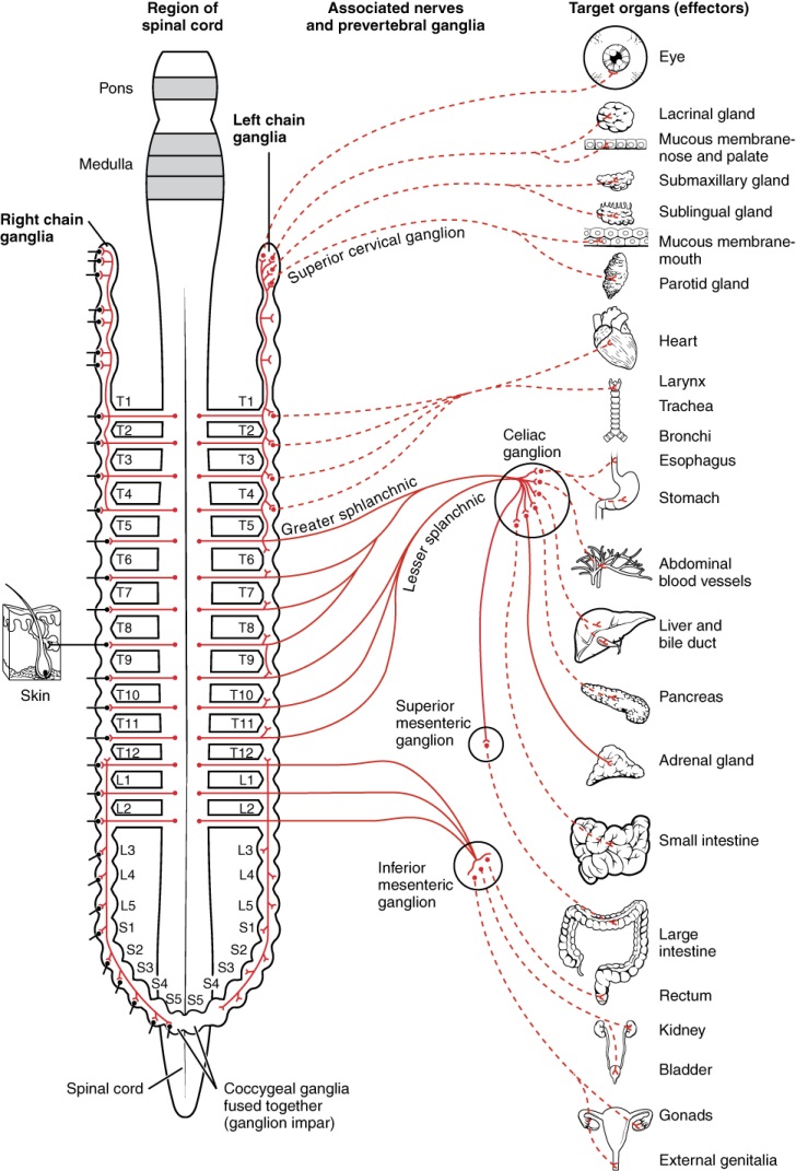
Figure 4: Neurons from the lateral horn of the spinal cord (preganglionic nerve fibers - solid lines)) project to the sympathetic chain ganglia on either side of the vertebral column or to collateral (prevertebral) ganglia that are anterior to the vertebral column in the abdominal cavity. Axons from these ganglionic neurons (postganglionic nerve fibers - dotted lines) then project to target effectors throughout the body.
Notice the length of the pre and postganglionic neurons differ because of the location of the autonomic ganglion to the spinal cord.
Not all axons from the central neurons terminate in the chain ganglia. Additional branches from the ventral nerve root continue through the chain and on to one of the collateral ganglia as the greater splanchnic nerve or lesser splanchnic nerve. For example, the greater splanchnic nerve at the level of T5 synapses with a collateral ganglion outside the chain before making the connection to the postganglionic nerves that innervate the stomach (see Figure 5c).
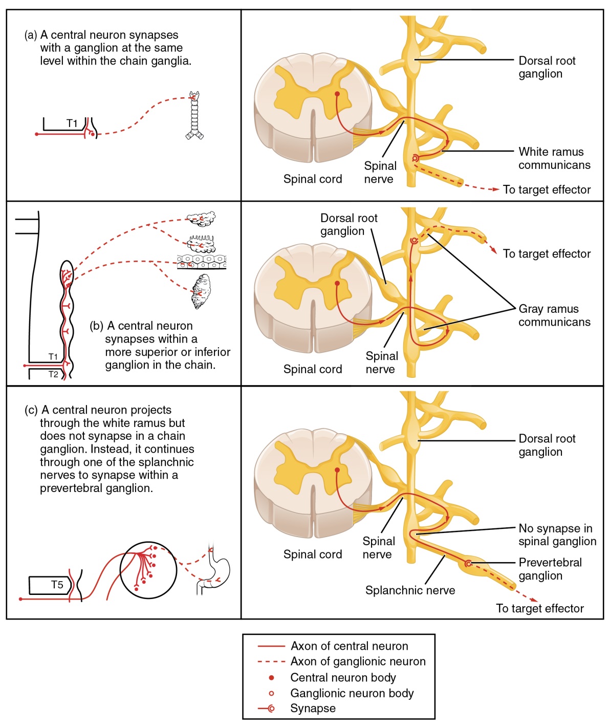
Figure 5: a) A central neuron synapses with a ganglion at the same level of the chain ganglia. b) A central neuron synapses within a more superior or inferior ganglion in the chain. c) The greater splanchnic nerve at the level of T5 synapses with a collateral ganglion outside the chain before making the connection to the postganglionic nerves that innervate the stomach. A splanchnic neuron is one that does not synapse within a chain ganglion, but continues to a prevertebral ganglion or to the effector where it connects with a postganglionic neuron.
