

Spinal Nerves
The nerves connected to the spinal cord are the spinal nerves. All of the spinal nerves are combined sensory and motor axons that separate into two nerve roots. The sensory axons enter the spinal cord as the dorsal nerve root. The motor fibers, both somatic and autonomic, emerge as the ventral nerve root. The dorsal root ganglion found only on the dorsal root (hence its name) is made of the large conglomerated neuronal cell bodies packed into the ganglion.
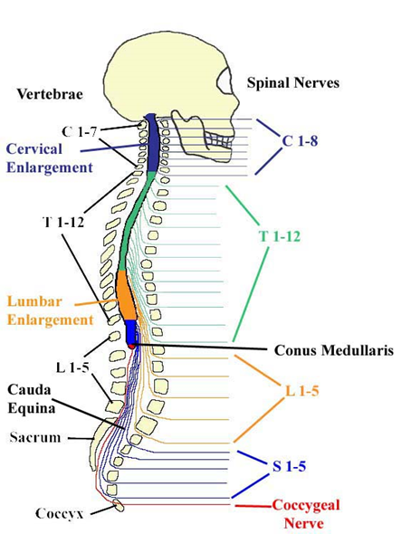
Figure 3. A skeleton view of the Spinal Cord, including where to find the 31 spinal nerves, the two spinal cord enlargements, and the location of the conus medullaris.
There are 31 spinal nerves, named for the level of the spinal cord at which each one emerges. There are eight pairs of cervical nerves designated C1 to C8, twelve thoracic nerves designated T1 to T12, five pairs of lumbar nerves designated L1 to L5, five pairs of sacral nerves designated S1 to S5, and one pair of coccygeal nerves. The nerves are numbered from the superior to inferior positions, and each emerges from the vertebral column through the intervertebral foramen at its level. The first nerve, C1, emerges between the first cervical vertebra and the occipital bone. The second nerve, C2, emerges between the first and second cervical vertebrae. The same occurs for C3 to C7, but C8 emerges between the seventh cervical vertebra and the first thoracic vertebra. For the thoracic and lumbar nerves, each one emerges between the vertebra that has the same designation and the next vertebra in the column. The sacral nerves emerge from the sacral foramina along the length of that unique vertebra.
Spinal nerves of the thoracic region, T2 through T11, are not part of the plexuses but rather emerge and give rise to the intercostal nerves found between the ribs, which articulate with the vertebrae surrounding the spinal nerve.
Spinal nerves extend outward from the vertebral column to innervate the periphery. The nerves in the periphery are not straight continuations of the spinal nerves, but rather the reorganization of the axons in those nerves to follow different courses. Axons from different spinal nerves will come together into a systemic nerve. This occurs at four places along the length of the vertebral column, each identified as a nerve plexus, whereas the other spinal nerves directly correspond to nerves at their respective levels. In this instance, the word plexus is used to describe networks of nerve fibers with no associated cell bodies.
Nerve Plexuses
There are four main nerve plexuses in the human body. The cervical plexus supplies nerves to the posterior head and neck, as well as the phrenic nerve to the diaphragm. The brachial plexus supplies five primary nerves to the arm - axillary, median, musculocutaneous, radial and ulnar nerves. The lumbar plexus supplies the femoral and other nerves to the anterior leg. The sacral plexus leads to the sciatic nerve and supplies nerves to the posterior leg. The location of the conus medularis lies about the area of L1 and L2, above the iliac crests.
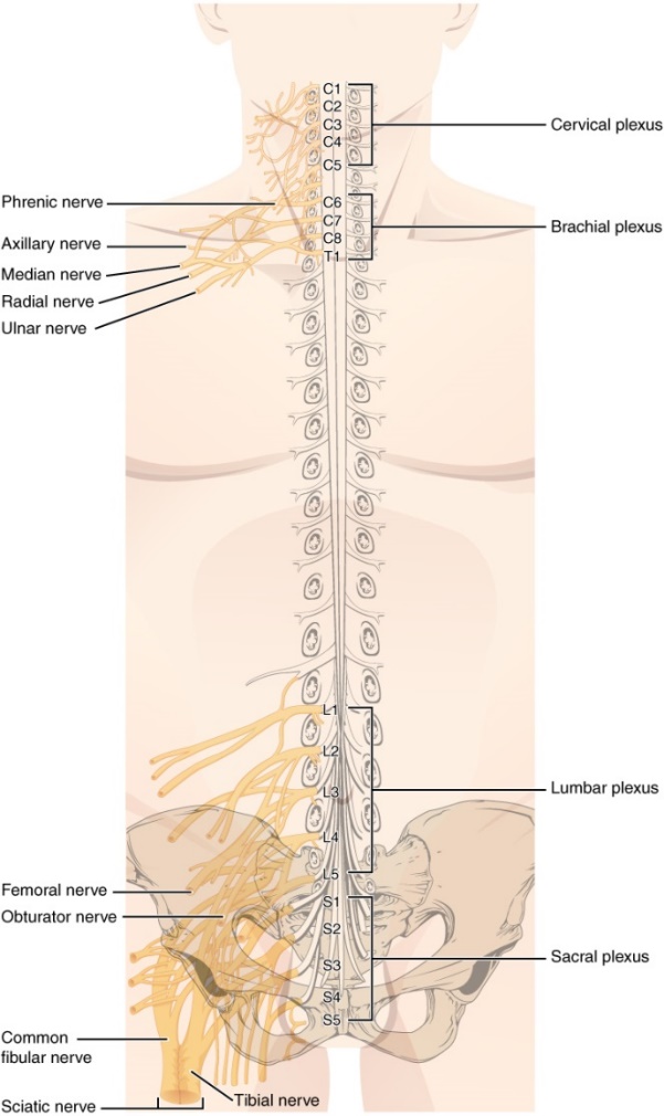
Figure 4 . There are four main nerve plexuses in the human body and 31 spinal nerves surrounded by the dura mater. The plexus networks are the cervical plexus, the brachial plexus, the lumbar plexus, and the sacral plexus (or the lumbosacral plexus). There are two enlargements, cervical enlargement and the lumbar enlargement, where a group of neuron cell bodies are located that supply the nerves to the large plexuses. Notice that the thoracic nerves do not have a plexus corresponding to them. Can you think of why?
{Response: There are ribs in the way so that the interconnections of a plexus network cannot be formed.}
Generally, spinal nerves contain afferent axons from sensory receptors in the periphery, such as from the skin, mixed with efferent axons travelling to the muscles or other effector organs. As the spinal nerve nears the spinal cord, it splits into dorsal and ventral roots. The dorsal root contains only the axons of sensory neurons, whereas the ventral roots contain only the axons of the motor neurons. Some of the branches will synapse with local neurons in the dorsal root ganglion, posterior (dorsal) horn, or even the anterior (ventral) horn, at the level of the spinal cord where they enter. Other branches will travel a short distance up or down the spine to interact with neurons at other levels of the spinal cord. A branch may also turn into the posterior (dorsal) column of the white matter to connect with the brain. For the sake of convenience, we will use the terms ventral and dorsal in reference to structures within the spinal cord that are part of these pathways. This will help to underscore the relationships between the different components. Typically, spinal nerve systems that connect to the brain are contralateral, in that the right side of the body is connected to the left side of the brain and the left side of the body to the right side of the brain.
