

Gray Matter and White Matter
Gray Horns
In cross-section, the gray matter of the spinal cord has the appearance of an ink-blot test, with the spread of the gray matter on one side replicated on the other—a shape reminiscent of a swollen capital "H." As shown in Figure 4, the gray matter is subdivided into regions that are referred to as horns. The posterior horn is responsible for sensory processing. The anterior horn sends out motor signals to the skeletal muscles. The lateral horn, which is only found in the thoracic, upper lumbar, and sacral regions, is the central component of the sympathetic division of the autonomic nervous system.
Some of the largest neurons of the spinal cord are the multipolar motor neurons in the anterior horn. The fibers that cause contraction of skeletal muscles are the axons of these neurons. The motor neuron that causes contraction of the big toe, for example, is located in the sacral spinal cord. The axon that has to reach all the way to the belly of that muscle may be a meter in length. The neuronal cell body maintaining that long axonal fiber is quite large, possibly several hundred micrometers in diameter, and makes it one of the largest cells in the body.
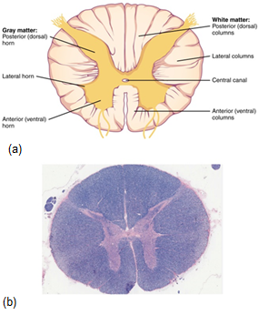
Figure 6. (a) The cross-section of a thoracic spinal cord segment shows the posterior, anterior, and lateral horns of gray matter, which make the shape of an "H" in the middle of the spinal cord. Also shown are the posterior, anterior, and lateral columns of white matter, which surround this letter "H". In the center of the gray matter ("H") is found the small Central Canal, which holds cerebrospinal fluid. LM × 40. (b) shows an actual micrograph provided by the Regents of University of Michigan Medical School © 2012
White Columns
Just as the gray matter is separated into horns, the white matter of the spinal cord is separated into columns. Ascending tracts of nervous system fibers in these columns carry sensory information up to the brain, whereas descending tracts carry motor commands from the brain. Looking at the spinal cord longitudinally, the column-like tracts extend along its length as continuous bands of white matter. Between the two posterior horns of gray matter are the posterior columns of white matter . Between the two anterior horns, and bounded by the axons of motor neurons emerging from that gray matter area, are the anterior columns. The white matter on either side of the spinal cord, between the posterior horn and the axons of the anterior horn neurons, are the lateral columns. The posterior columns are composed of axons of ascending tracts. The anterior and lateral columns are composed of many different groups of axons of both ascending and descending tracts — the latter carrying motor commands down from the brain to the spinal cord to control output to the periphery.
Also, remember the anterior midline is marked by the anterior median fissure, and the posterior midline is marked by the posterior median sulcus. Can you find these on the figure below?
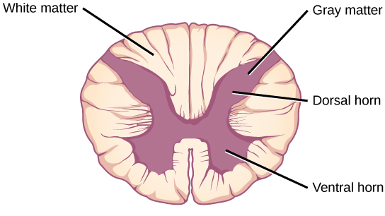
Figure 7. A cross-section of the spinal cord shows gray matter (containing cell bodies and unmyelinated interneurons) and white matter (containing myelinated axons).
Video

