Origin, Structure and Functions of the Cranial Nerves and All Parts of the Brain
Anatomy & Physiology I
Origin, Structure and Functions of the Cranial Nerves and All Parts of the Brain
Objectives:
By the end of this section, the student will be able to:
- Relate the different stages of development to the adult structures of the central nervous system
- Locate regions of the cerebral cortex by common anatomical landmarks
- Relate the structures of the CNS with their functions
- Describe the protective coverings of the CNS and how they protect the brain and spinal cord
- Explain the blood-brain barrier and how the blood and cerebrospinal fluid nourish and protect the brain
- Explain the expansion of the ventricular system of the adult brain from the central canal of the neural tube
- Describe the connections of the diencephalon and cerebellum on the basis of patterns of embryonic development
- Understand the blood circulation to the brain is provided by two pathways into a loop called the Circle of Willis
The brain is a complex organ composed of gray parts and white matter, which can be hard to distinguish. Starting from an embryologic perspective allows you to understand more easily how the parts relate to each other. The embryonic nervous system begins as a very simple structure—essentially just a straight line, which then gets increasingly complex. Looking at the development of the nervous system with a couple of early snapshots makes it easier to understand the whole complex system. By following the developmental pattern, it is possible to learn what the major regions of the nervous system are, their locations and functions. There is a physiological concept known as localization of function that states that certain structures are specifically responsible for prescribed functions. It is an underlying concept in all of anatomy and physiology, but the nervous system illustrates the concept very well.

Figure 1. Gray Matter and White Matter. A brain removed during an autopsy, with a partial section removed, shows white matter surrounded by gray matter on the surface. Gray matter makes up the outer layer or cortex of the brain.
(credit: modification of work by "Suseno"/Wikimedia Commons)
The brain develops from the ectoderm layer of the growing embryo and forms a neural tube. Before neural tube is complete, the cephalic portion starts to grow faster. This enlarged portion starts to grow into three vesicles: the prosencephalon (prō-zen-SEF-a-lon), or forebrain (pro- means "before," but also think of a "pro" athlete always in the front); the mesencephalon, or midbrain; and the rhombencephalon (rom-ben-SEF-a-lon), or hindbrain (think of the "rump" or your backend to remember the hindbrain). Except for the midbrain, or mesencephalon, the vesicles will divide into more vesicles and then into the parts of the brain during development. The "pros" becomes the cerebrum and diencephalon (the forefront of our thinking and homeostasis), the "mes" remains the midbrain, and the "rhomb" becomes the inferior and posterior portions of the brain stem and cerebellum (Figure 2).
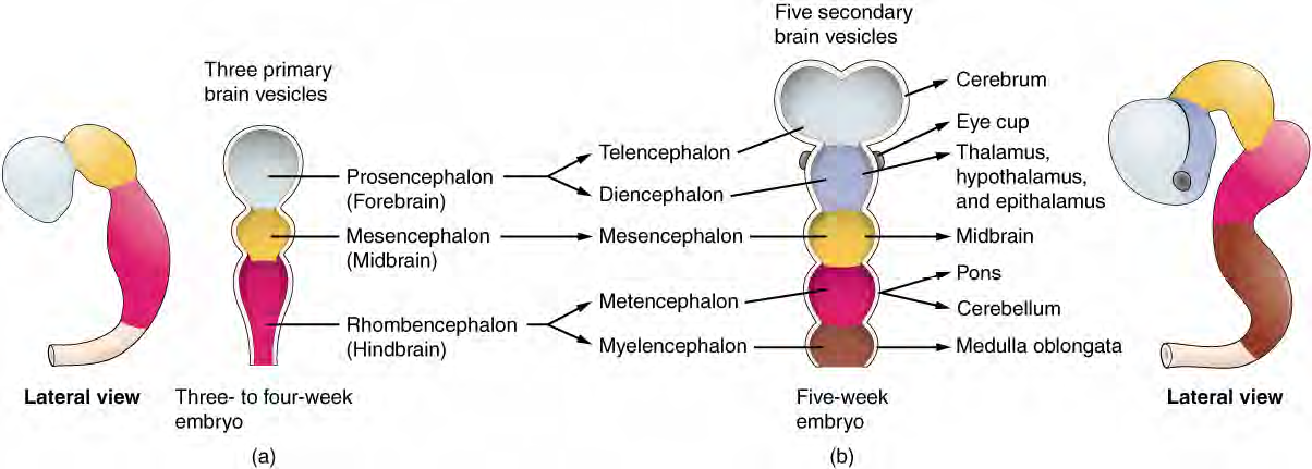
Figure 2. Development of the Brain includes three primary brain vesicles, the Prosencephalon (forebrain), Mesencephalon (midbrain), Rhombencephalon (hindbrain). These develop into the primary parts of the brain - Cerebrum, Diencephalon, Midbrain, Pons, Cerebellum, and the Medulla oblongata.
The early nervous system is a simple hollow tube that runs from the anterior end of the embryo to the posterior end. Beginning at 25 days, the anterior end develops into the brain, and the posterior portion becomes the spinal cord. This is the most basic arrangement of tissue in the nervous system, and it gives rise to the more complex structures by the fourth week of development.
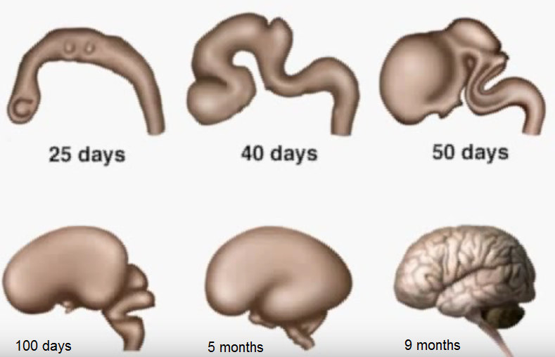
Figure 3. Human Fetal Brain Development Milestones at 25 days, 40 days, 50 days, 100 days, 5 months and 9 months.
We can see various connections within the embryonic brain when we later examine the special senses. For instance, the retina, which began as part of the diencephalon, is primarily connected to the diencephalon. The eyes are just inferior to the anterior-most part of the cerebrum, but the optic nerve extends back to the thalamus as the optic tract, with branches into a region of the hypothalamus. There is also a connection of the optic tract to the midbrain, but the mesencephalon is adjacent to the diencephalon, so that is not difficult to imagine.
We know that the brain develops over childhood through adulthood. If the brain (especially the prefrontal cortex) does not finish developing until approximately 20 years of age, can teenagers be held responsible for behaving badly? Read on about the anatomy of the brain, recalling its development as you go.

The Cerebrum
The iconic gray mantle of the human brain, which appears to make up most of the mass of the brain, is the cerebrum ( Figure below ). The wrinkled portion is the cerebral cortex , and the rest of the structure is beneath that outer covering. There is a large separation between the two sides of the cerebrum called the longitudinal fissure. It separates the cerebrum into two distinct halves, a right and left cerebral hemisphere . Deep within the cerebrum, the white matter of the corpus callosum provides the major pathway for communication between the two hemispheres of the cerebral cortex.

Figure 4. Anterior and Lateral views of the Brain. The outmost layer of the cerebrum is the cerebral cortex. The cerebrum is divided into right and left hemispheres. The corpus callosum joins the two sides with bundles of myelinated axons.
Many of the higher neurological functions, such as memory, emotion, and consciousness, are the result of cerebral function. The complexity of the cerebrum is different across vertebrate species. The cerebrum of the most primitive vertebrates is not much more than the connection for the sense of smell. In mammals, the cerebrum comprises the outer gray matter that is the cortex (from the Latin word meaning "bark of a tree") and several deep nuclei that belong to three important functional groups. The basal nuclei are responsible for cognitive processing, the most important function being that associated with planning movements. The basa forebrain contains nuclei that are important in learning and memory. The limbic cortex is the region of the cerebral cortex that is part of the limbic system, a collection of structures involved in emotion, memory, and behavior.
Cerebral Cortex
The cerebrum is covered by a continuous layer of gray matter that wraps around either side of the forebrain—the cerebral cortex. This thin, extensive region of wrinkled gray matter is responsible for the higher functions of the nervous system. A gyrus (plural = gyri) is the ridge of one of those wrinkles, and a sulcus (plural = sulci) is the groove between two gyri. The pattern of these folds of tissue indicates specific regions of the cerebral cortex. The head is limited by the size of the birth canal, and the brain must fit inside the cranial cavity of the skull. Extensive folding in the cerebral cortex enables more gray matter to fit into this limited space. If the gray matter of the cortex were peeled off of the cerebrum and laid out flat, its surface area would be roughly equal to one square meter. The folding of the cortex maximizes the amount of gray matter in the cranial cavity. During embryonic development, as the telencephalon expands within the skull, the brain goes through a regular course of growth that results in everyone's brain having a similar pattern of folds. The surface of the brain can be mapped on the basis of the locations of large gyri and sulci.
Using these landmarks, the cortex can be separated into four major regions, or lobes (Figure 5). The lateral sulcus that separates the temporal lobe from the other regions is one such landmark. Superior to the lateral sulcus are the parietal lobe and frontal lobe, which are separated from each other by the central sulcus. The posterior region of the cortex is the occipital lobe, which has no obvious anatomical border between it and the parietal or temporal lobes on the lateral surface of the brain. From the medial surface, an obvious landmark separating the parietal and occipital lobes is called the parieto-occipital sulcus. The fact that there is no obvious anatomical border between these lobes is consistent with the functions of these regions being interrelated.
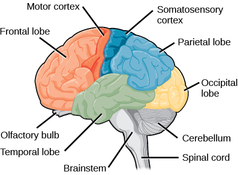
Figure 5. Parts of the brain. Frontal lobe, parietal lobe, occipital lobe, temporal lobe, cerebellum, and brain stem are shown in this figure.
Different regions of the cerebral cortex can be associated with particular functions, a concept known as localization of function. The temporal lobe is associated with primary auditory sensation, known as Brodmann's areas 41 and 42 in the superior temporal lobe. Because regions of the temporal lobe are part of the limbic system, memory is an important function associated with that lobe. Memory is essentially a sensory function; memories are recalled sensations such as the smell of Mom's baking or the sound of a barking dog. Even memories of movement are really the memory of sensory feedback from those movements, such as stretching muscles or the movement of the skin around a joint. Structures in the temporal lobe are responsible for establishing long-term memory, but the ultimate location of those memories is usually in the region in which the sensory perception was processed.
The main sensation associated with the parietal lobe is somatosensation, meaning the general sensations associated with the body. Posterior to the central sulcus is the postcentral gyrus, the primary somatosensory cortex, which is identified as Brodmann's areas 1, 2, and 3. All of the tactile senses are processed in this area, including touch, pressure, tickle, pain, itch, and vibration, as well as more general senses of the body such as proprioception and kinesthesia, which are the senses of body position and movement, respectively.
Anterior to the central sulcus is the frontal lobe, which is primarily associated with motor functions. The precentral gyrus is the primary motor cortex. Cells from this region of the cerebral cortex are the upper motor neurons that instruct cells in the spinal cord to move skeletal muscles. Anterior to this region are a few areas that are associated with planned movements. The premotor area is responsible for thinking of a movement to be made. The frontal eye fields are important in eliciting eye movements and in attending to visual stimuli. Broca's area is responsible for the production of language, or controlling movements responsible for speech; in the vast majority of people, it is located only on the left side. Anterior to these regions lies the prefrontal lobe, which serves cognitive functions that can be the basis of personality, decision-making, understanding consequences, and consciousness. The prefrontal lobotomy is an outdated mode of treatment for personality disorders (psychiatric conditions) that profoundly affected the personality of the patient. You may think that your teenager has had a prefrontal lobotomy with the lack of good decision-making she may show. However, under the age of 25 the prefrontal lobe has not completely developed for all young people. A lot of instinctual decisions and quick reactions to situations or aggressive behavior come from the emotional area of the brain called the amygdala which develops early. So a teenager's or young adult's decisions are based more on this emotional brain rather than the logical brain.
Phineas P. Gage
(1823 - May 21, 1860) was an American railroad construction foreman remembered for his improbable survival of an accident in which a large iron rod was driven completely through his head, destroying much of his brain's left frontal lobe , and for that injury's reported effects on his personality and behavior over the remaining twelve years of his life—effects so profound that (for a time at least) friends saw him as "no longer Gage."
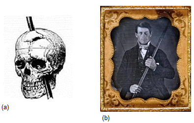
Figure 6. Phineas Gage, the iron's path (b) is from the collection of Jack and Beverly Wilgus.
Phineas Gage influenced nineteenth-century discussion about the mind and brain, particularly debate on cerebral localization , and was perhaps the first case to suggest that damage to specific parts of the brain might induce specific personality changes.
A report of Gage's physical and mental condition shortly before his death implies that his most serious mental changes were temporary, so that in later life he was far more functional, and socially far better adapted, than in the years immediately following his accident. A social recovery hypothesis suggests that his employment as a stagecoach driver in Chile provided daily structure allowing him to regain some lost social and personal skills. All of this points to the remarkableness of the brain, the psychology of it, and its recoverability.
How Much of Your Brain Do You Use?
Have you ever heard the claim that humans only use 10 percent of their brains? Maybe you have seen an advertisement on a website saying that there is a secret to unlocking the full potential of your mind—as if there were 90 percent of your brain sitting idle, just waiting for you to use it. If you see an ad like that, don't click. It isn't true!
An easy way to see how much of the brain a person uses is to take measurements of brain activity while performing a task. An example of this kind of measurement is functional magnetic resonance imaging (fMRI), which generates a map of the most active areas and can be generated and presented in three dimensions (Figure 7). This procedure is different from the standard MRI technique because it is measuring changes in the tissue in time with an experimental condition or event.
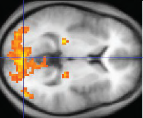
Figure 7. fMRI. This functional MRI shows activation of the visual cortex in response to visual stimuli.
(credit: "Superborsuk"/Wikimedia Commons)
Subcortical Structures
The hippocampus and amygdala are medial-lobe structures that, along with the adjacent cortex, are involved in long-term memory formation and emotional responses. The basal nuclei are a set of nuclei in the cerebrum responsible for comparing cortical processing with the general state of activity in the nervous system to influence the likelihood of movement taking place. For example, while a student is sitting in a classroom listening to a lecture, the basal nuclei will keep the urge to jump up and scream from actually happening.
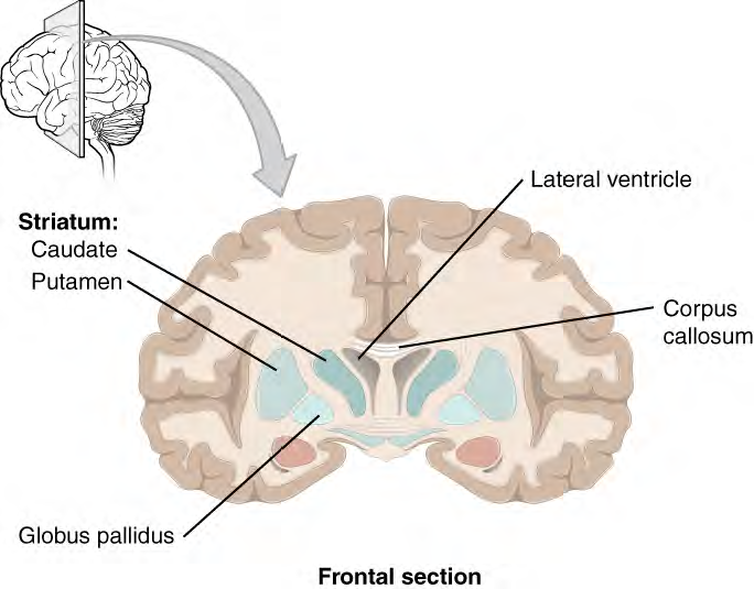
Figure 8 . Frontal Section of Cerebral Cortex and Basal Nuclei The major components of the basal nuclei, shown in a frontal section of the brain, are the caudate (just lateral to the lateral ventricle), the putamen (inferior to the caudate and separated by the large white-matter structure called the internal capsule), and the globus pallidus (medial to the putamen).
Disorders of the Basal Nuclei
Parkinson's disease is a disorder of the basal nuclei, specifically of the substantia nigra, which releases dopamine. Parkinson's disease is the result of neurons in the substantia nigra pars compacta pathway dying. The reduction of signal through this pathway results in the tremor and other symptoms of the disease.
Parkinson's disease is neurodegenerative, meaning that neurons die that cannot be replaced, so there is no cure for the disorder. Treatments for Parkinson's disease are aimed at increasing dopamine levels in the striatum. Currently, the most common way of doing that is by providing the amino acid L-DOPA, which is a precursor to the neurotransmitter dopamine and can cross the blood-brain barrier. With levels of the precursor elevated, the remaining cells of the substantia nigra pars compacta can make more neurotransmitter and have a greater effect. Unfortunately, the patient will become less responsive to L-DOPA treatment as time progresses, and it can cause increased dopamine levels elsewhere in the brain, which are associated with psychosis or schizophrenia.
View the information on Parkinson Disease (opens in new window) for a more thorough explanation of Parkinson's disease.
The Myth of Left Brain/Right Brain
There is a persistent myth that people are "right-brained" or "left-brained," which is an oversimplification of an important concept about the cerebral hemispheres. There is some lateralization of function, in which the left side of the brain is devoted to language function and the right side is devoted to spatial and nonverbal reasoning. Whereas these functions are predominantly associated with those sides of the brain, there is no monopoly by either side on these functions. Many pervasive functions, such as language, are distributed globally around the cerebrum.
Some of the support for this misconception has come from studies of split brains. A drastic way to deal with a rare and devastating neurological condition (intractable epilepsy) is to separate the two hemispheres of the brain. After sectioning the corpus callosum, a split-brained patient will have trouble producing verbal responses on the basis of sensory information processed on the right side of the cerebrum, leading to the idea that the left side is responsible for language function.
However, there are well-documented cases of language functions lost from damage to the right side of the brain. The deficits seen in damage to the left side of the brain are classified as aphasia, a loss of speech function; damage on the right side can affect the use of language. Right-side damage can result in a loss of ability to understand figurative aspects of speech, such as jokes, irony, or metaphors. Nonverbal aspects of speech can be affected by damage to the right side, such as facial expression or body language, and right-side damage can lead to a "flat affect" in speech, or a loss of emotional expression in speech—sounding like a robot when talking.
The Diencephalon (di-en-SEF-a-lon)
The diencephalon is the connection between the cerebrum and the rest of the nervous system, with one exception. The rest of the brain, the spinal cord, and the PNS all send information to the cerebrum through the diencephalon. Output from the cerebrum passes through the diencephalon. The single exception is the system associated with olfaction, or the sense of smell, which connects directly with the cerebrum.
The diencephalon is deep beneath the cerebrum and makes up the walls of the third ventricle. The diencephalon can be described as any region of the brain with "thalamus" in its name. The two major regions of the diencephalon are the thalamus itself and the hypothalamus (see figure below). There are other structures, such as the epithalamus, which contains the pineal (`pin-ē-al) gland, or the subthalamus, which includes the subthalamic nucleus that is part of the basal nuclei.
Thalamus
The thalamus is a collection of nuclei that relay information between the cerebral cortex and the PNS, spinal cord, or brain stem. All sensory information, except for the sense of smell, passes through the thalamus before processing by the cortex. Axons from the peripheral sensory organs (first order neuron) synapse in the thalamus, and thalamic neurons (second order neuron) project directly to the cerebrum. The thalamus acts like a relay station or a receptionist asking "How may I direct your call?" and sending the incoming information from the first order neuron to the correct location on the cortex via the second order neuron. The thalamus does not just pass the information on, it also processes that information. For example, the portion of the thalamus that receives visual information will influence what visual stimuli are important, or what receives attention.
The cerebrum also sends information down to the thalamus, which usually communicates motor commands. This involves interactions with the cerebellum and other nuclei in the brain stem. The cerebrum interacts with the basal nuclei, which involves connections with the thalamus.
Hypothalamus
Inferior and slightly anterior to the thalamus is the hypothalamus, the other major region of the diencephalon. The hypothalamus is a collection of nuclei that are largely involved in regulating homeostasis. The hypothalamus is the executive region in charge of the autonomic nervous system and the endocrine system through its regulation of the anterior pituitary gland. Other parts of the hypothalamus are involved in memory and emotion as part of the limbic system.

Figure 9. The diencephalon is composed primarily of the thalamus and hypothalamus , which together define the walls of the third ventricle. The thalami are two elongated, ovoid structures ("bird's head") on either side of the midline that make contact in the middle. The hypothalamus is inferior and anterior to the thalamus, culminating in a beak-like sharp angle to which the pituitary gland is attached. Above the diencephalon, the corpus callosum and the fornix can be seen (similar to crests on a bird's head).
The thalami are two elongated, ovoid structures on either side of the midline that make contact in the middle - at the "eye" of the bird's head called the Intermediate Mass of the thalamus. The hypothalamus is inferior and anterior to the thalamus, culminating in a "beak-like" structure to which the pituitary gland is attached.
The limbic system is a connected set of structures between the forebrain and hindbrain that regulates emotion, as well as behaviors related to fear and motivation. It plays a role in memory formation and includes parts of the thalamus and hypothalamus as well as the hippocampus. One important structure within the limbic system is a temporal lobe structure called the amygdala, mentioned earlier . The two amygdala (one on each side) are important both for the sensation of fear and for recognizing fearful faces. As aforementioned the amygdala develops early in life and persists as the lead emotional decision-maker until the prefrontal cortex develops fully in the mid-20s.
Brain Disorders
- Schizophrenia is characterized by psychotic episodes, hallucinations, delusions
- there may be multiple genes responsible for it
- treatment focuses on the use of dopamine
- amphetamine, which stimulates dopamine release
- drugs that alleviate symptoms of schizophrenia block dopamine receptors
- other neurotransmitters affected are serotonin and norepinephrine, and glutamate
- PCP blocks glutamate receptors and produces schizophrenia like symptoms
- Depressive illnesses are generally bipolar and major depression
- bipolar disorders involve mood swings from very high to very low
- in the manic phase, increased energy, risky behaviors, high self-esteem occur
- in the depressive phase, feelings of worthlessness, sleep disturbances, loss of interest, and risk of suicide occur
- those with major depression experience a low mood most of the time
- treatments include Prozac, electroconvulsive shock, lithium and talk therapy
- Alzheimer's disease
- is caused by mental deterioration which leads to dementia
- many symptoms occur, including confusion, memory loss, disorientation, and more
- diagnosis is usually made on autopsy, finding huge areas of the brain that have died or shrunk
- neurons have neurofibrillary tangles and senile plaques
- neurofibrillary tangles are bundles of degenerated neuron and glial processes
- senile plaques are clusters of beta-amyloid protein, cleaved from a normal protein found in healthy neurons
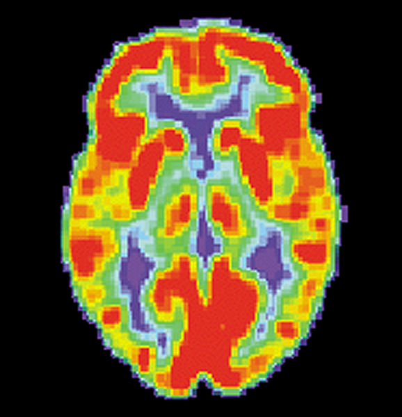
Figure 10. Normal Brain PET Scan. By US National Institute on Aging, Alzheimer's Disease Education and Referral Center
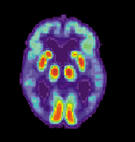
Figure 11. Alzheimer Brain PET Scan. Please compare the orange levels of activity seen in this brain and the normal brain in Figure 10. By US National Institute on Aging, Alzheimer's Disease Education and Referral Center
- Parkinson's disease
- characterized by difficulty in smooth movement, with muscle rigidity near death
- is caused by neuron death in the midbrain nucleus, called the substantia nigra, which release dopamine
- there is no known cause of the disease, probably is due to a combination of environmental and hereditary factors
- L-dopa has served as a treatment
- embryonic stem cell treatment may provide a cure in the future
Brain Stem
The midbrain and hindbrain (composed of the pons and the medulla) are collectively referred to as the brain stem. The structure emerges from the ventral surface as a cone that connects the brain to the spinal cord. The midbrain coordinates sensory representations of the visual, auditory, and somatosensory perceptual spaces. The pons is the main connection with the adjacent cerebellum. The pons and the medulla regulate several crucial functions, including the cardiovascular and respiratory systems and rates.
The cranial nerves connect through the brain stem and provide the brain with the sensory input and motor output associated with the head and neck, including most of the special senses. The major ascending and descending pathways between the spinal cord and brain, specifically the cerebrum, pass through the brain stem.
Midbrain
One of the original regions of the embryonic brain, the midbrain is a small region between the thalamus and pons. It is separated into the tectum and tegmentum, from the Latin words for roof and floor, respectively. The tectum is composed of four bumps known as the colliculi (singular = colliculus), which means "little hill". The inferior colliculus is the inferior pair of these enlargements and is part of the auditory brain stem pathway. Neurons of the inferior colliculus project to the thalamus, which then sends auditory information to the cerebrum for the conscious perception of sound. Fibers between the inferior and the superior colliculi give auditory input that stimulates visual reflexes to a loud sound, etc.
The superior colliculus involved with visual reflexes combining sensory information about visual space, auditory space, and somatosensory space. Activity in the superior colliculus is related to orienting the eyes to a vision, sound or touch stimulus. If you are walking along the sidewalk on campus and you hear chirping, the superior colliculus coordinates that information with your awareness of the visual location of the tree right above you. That is the correlation of auditory and visual maps. If you suddenly feel something wet fall on your head, your superior colliculus integrates that with the auditory and visual maps and you know that the chirping bird just relieved itself on you. You want to look up to see the culprit, but do not!
The tegmentum is continuous with the gray matter of the rest of the brain stem. Throughout the midbrain, pons, and medulla, the tegmentum contains the nuclei that receive and send information through the cranial nerves, as well as regions that regulate important functions such as those of the cardiovascular and respiratory systems.
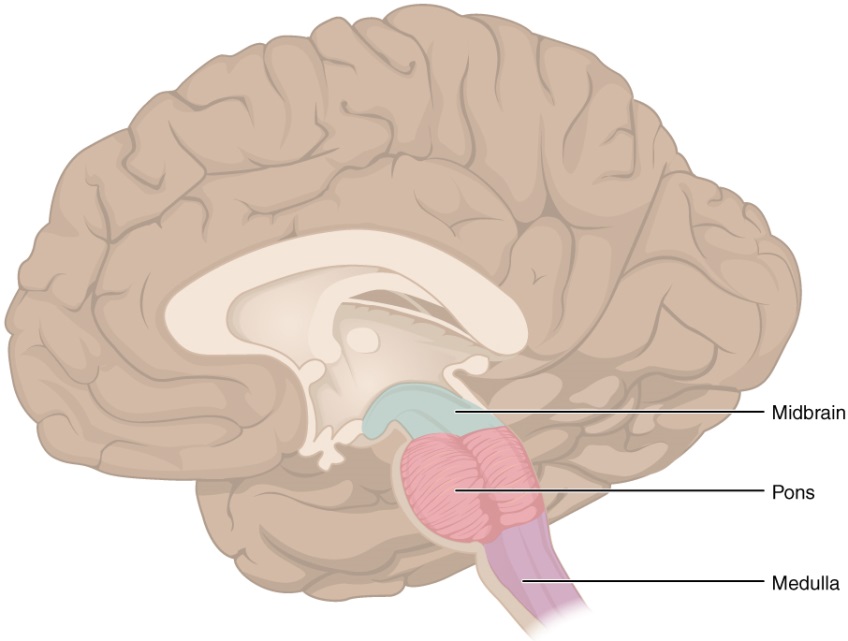
Figure 12. The brain stem comprises three regions: the midbrain, the pons and the medulla.
Pons
The word pons comes from the Latin word for bridge. It is visible on the anterior surface of the brain stem as the thick bundle of white matter attached to the cerebellum. The pons is the main connection between the cerebellum and the brain stem. The bridge-like white matter looks like a little pooch on the anterior side of the brain stem area. Gray matter in the interior (tegmentum region) of the pons contains neurons receiving descending (motor) input from the forebrain that is sent to the cerebellum, mostly to coordinate movements.
Medulla (me'-dull-a)
The medulla is the region known as the myelencephalon in the embryonic brain. The initial portion of the name, "myel," refers to the abundant white matter found here, which is continuous with the white matter of the spinal cord. The tegmentum of the midbrain and pons continues into the medulla because this gray matter is responsible for processing cranial nerve information. A diffuse region of gray matter throughout the brain stem, known as the reticular formation, is related to sleep and wakefulness, such as general brain activity and attention, especially to stay awake during long lectures.
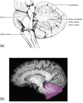
Figure 13. The cerebellum is situated on the posterior surface of the brain stem. Descending input from the cerebellum enters through the large white matter structure of the pons. Ascending input from the periphery and spinal cord enters through the fibers of the inferior olive. Output goes to the midbrain, which sends a descending signal to the spinal cord.
The Cerebellum
The cerebellum, as the name suggests, is the "little brain." It is covered in gyri and sulci like the cerebrum, and looks like a miniature version of that part of the brain (Figure above). The cerebellum is largely responsible for comparing information from the cerebrum with sensory feedback from the periphery through the spinal cord. So it is responsible for coordinating the movements of the body and it accounts for about 10 percent of the brain.
Descending fibers from the cerebrum have branches that connect to neurons in the pons. Those neurons project into the cerebellum, providing a copy of motor commands sent to the spinal cord. If the primary motor cortex of the frontal lobe sends a command down to the spinal cord to initiate walking, a copy of that instruction is sent to the cerebellum. Sensory feedback from the muscles and joints, proprioceptive (position in space) information about the movements of walking, and sensations of balance are sent to the cerebellum, which will compare them. If walking is not coordinated, perhaps because the ground is uneven or a strong wind is blowing, then the cerebellum sends out a corrective command to compensate for the difference between the original cortical command and the sensory feedback. The output of the cerebellum is into the midbrain, which then sends a descending input to the spinal cord to correct the messages going to skeletal muscles for the coordination and balance needed.
Meninges
The outer surface of the CNS is covered by a series of membranes composed of dense, irregular connective tissue called the meninges, which protect the brain. The dura mater is a thick fibrous layer and a strong protective sheath over the entire brain and spinal cord. The dura mater has two fibrous layers (meningeal layer and the periosteal layer) enclosing dural sinuses, which carry blood from the brain to the internal jugular veins. (Sinuses are thinner-walled veins.) Three extensions of the dura mater separate parts of the brain. The falx cerebri, dives into longitudinal fissure to separate and stabilize cerebral hemispheres and keep them from colliding. The tentorium cerebelli is another fold of dura mater in the transverse fissure. It protects the cerebellum from the weight of the occipital lobes. The tentorium contours to the shape of the cerebri, and has the appearance of a 'tent' over the cerebellum. The falx cerebelli is a smaller portion of dura mater that separates the cerebellar hemispheres.
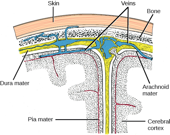
Figure 14. The cerebral cortex is covered by three layers of meninges: the dura, arachnoid, and pia maters. The yellow represents the dura that also surrounds the veins or dura sinus (blue). The arachnoid mater has a web-like appearance to it and extensions (granulations/arachnoid villi) that allow the cerebrospinal fluid to drain into the sinus (blue) and be added back to the blood stream. The thin pia mater (pink) lines the outside of the cerebral cortex like an onion skin.
The second meninx is the arachnoid mater, getting its name from the web-like extensions that go into the subarachnoid space. There is a thin subdural space between the dura mater and arachnoid mater that holds interstitial fluid. If you've heard of a subdural hematoma, it is an accumulation of blood outside of the brain in this space. However, the increase of pressure on the brain due to the increased blood causes symptoms to arise.
The outer surface of the brain and spinal cord is covered in the thin fibrous membrane of the pia mater. It is thought to have a continuous layer of cells providing a fluid-impermeable membrane. The pia mater extends across all gyri and into all sulci of the brain and cerebellar cortices. Because the spinal cord does not extend through the lower lumbar region of the vertebral column (find where it stops in the lumbar region on models and pictures), a needle can be inserted through the dura and arachnoid layers to withdraw CSF. This procedure is called a lumbar puncture.
Disorders of the Meninges
Meningitis is an inflammation of the meninges. Meningitis can be caused by infection by bacteria or viruses. The particular pathogens are not special to meningitis; it is just an inflammation of that specific set of tissues from what might be a broader infection. Bacterial meningitis can be caused by Streptococcus, Staphylococcus, or the tuberculosis pathogen, among many others. Viral meningitis is usually the result of common enteroviruses (such as those that cause intestinal disorders), but may be the result of the herpes virus or West Nile virus. Bacterial meningitis tends to be more severe.
The symptoms associated with meningitis can be fever, chills, nausea, vomiting, light sensitivity, soreness of the neck, or severe headache. A serious risk of meningitis can be damage to peripheral structures because of the nerves that pass through the meninges. Hearing loss is a common result of meningitis.
The primary test for meningitis is a lumbar puncture. Fatality occurs in approximately 22% of children, but more frequently in adults with bacterial meningitis. Treatment of bacterial meningitis is through antibiotics, but viral meningitis cannot be treated with antibiotics because viruses do not respond to that type of drug. Fortunately, the viral forms are milder.
Circulation for the Central Nervous System
This will be studied in more detail later, but we bring it to your attention here so you can understand when blood flow is blocked to the brain, there are other pathways feeding the brain.
Objectives:
By the end of this section, you will be able to:
- Describe the vessels that supply the CNS with blood
- Name the components of the ventricular system and the regions of the brain in which each is located
- Explain the production of cerebrospinal fluid and its flow through the ventricles
- Explain how a disruption in circulation would result in a stroke
The CNS is crucial to the operation of the body, and any compromise in the brain and spinal cord can lead to severe difficulties. The CNS has a privileged blood supply, as suggested by the blood-brain barrier. The function of the tissue in the CNS is crucial to the survival of the organism, so the contents of the blood cannot simply pass into the central nervous tissue. To protect this region from the toxins and pathogens that may be traveling through the blood stream, there is strict control over what can move out of the general systems and into the brain and spinal cord. Because of this privilege, the CNS needs specialized structures for the maintenance of circulation and a constant supply of fresh blood for glucose. This begins with a unique arrangement of blood vessels carrying fresh blood into the CNS. Beyond the supply of blood, the CNS filters that blood into cerebrospinal fluid (CSF), which is then circulated through the cavities of the brain and spinal cord called ventricles.
Blood Supply to the Brain
You will find all of these pathways and vessels in lab; you will also revisit this in the blood vessels module in the cardiovascular system later.
A lack of oxygen to the CNS can be devastating, and the cardiovascular system has specific regulatory reflexes to ensure that the blood supply is not interrupted. There are multiple routes for blood to get into the CNS, with specializations to protect that blood supply and to maximize the ability of the brain to get an uninterrupted perfusion.
Arterial Supply
The major artery carrying recently oxygenated blood away from the heart is the aorta. The very first branches off the aorta supply the heart with nutrients and oxygen. The next branches give rise to the common carotid arteries, which further branch into the internal carotid arteries. The external carotid arteries supply blood to the tissues on the surface of the cranium. The bases of the common carotids contain stretch receptors that immediately respond to the drop in blood pressure upon standing. The orthostatic reflex is a reaction to this change in body position, so that blood pressure is maintained against the increasing effect of gravity (orthostatic means "standing up"). Heart rate increases—a reflex of the sympathetic division of the autonomic nervous system— and this raises blood pressure.
The internal carotid artery enters the cranium through the carotid canal in the temporal bone. A second set of vessels that supply the CNS are the vertebral arteries, which are protected as they pass through the neck region by the transverse foramina of the cervical vertebrae. The vertebral arteries enter the cranium through the foramen magnum of the occipital bone. Branches off the left and right vertebral arteries merge into the anterior spinal artery supplying the anterior aspect of the spinal cord, found along the anterior median fissure. The two vertebral arteries then merge into the basilar artery, which gives rise to branches to the brain stem and cerebellum. The left and right internal carotid arteries and branches of the basilar artery all become the circle of Willis, a confluence of arteries that can maintain perfusion of the brain even if narrowing or a blockage limits flow through one part.
The figure below almost looks like a space alien. Imagine that the vertebral arteries are the alien's legs. Then the basilar artery is the body (also think of the ending of basil ar to remember that this is the artery's name, so as not to mix it up with a similar named vein). The posterior cerebral arteries could be its arms, while the middle and anterior arteries antennae, and the internal carotid artery openings are the alien's eyes.
Practice: Draw a picture of the "alien" yourself, with just these structures and label it. Do this several times until you get the blood vessels correct from memory.
Circle of Willis
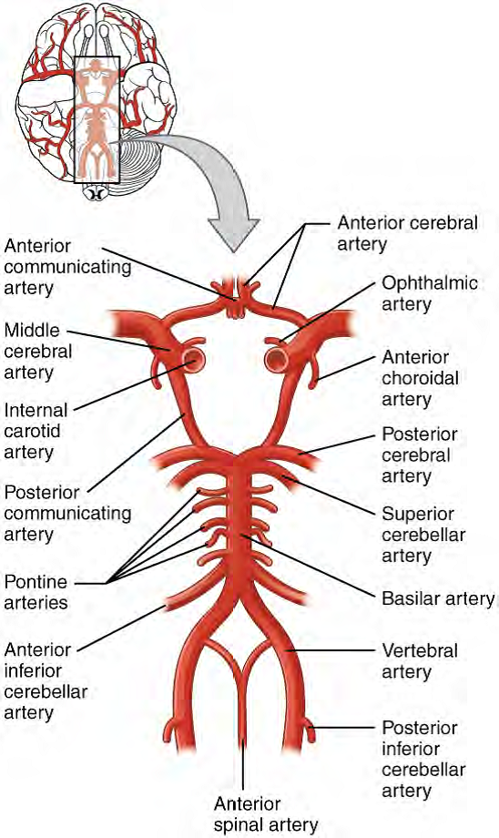
Figure 15. Circle of Willis. The blood supply to the brain enters through the internal carotid arteries and the vertebral arteries, up through the basilar artery, eventually giving rise to the circle of Willis. This arterial blood supply to the brain includes the following arteries seen in the figure: posterior cerebral artery, posterior communicating artery, middle cerebral artery (not part of actual Circle, though), anterior communicating artery and the anterior cerebral artery.
Venous Return
After passing through the CNS, blood returns to the circulation through a series of dural sinuses and veins mentioned before. The superior sagittal sinus runs in the groove of the longitudinal fissure, where it absorbs CSF from the meninges.

Figure 16. Dural Sinuses and Veins Blood drains from the brain through a series of sinuses that connect to the jugular veins. You will need to learn these for the Blood vessels module later.
Ventricles (sacs of fluid)
Another aspect of the adult CNS structures that relates to embryonic development is the ventricles—open spaces within the CNS where cerebrospinal fluid is produced and circulates. They are the remnant of the hollow center of the neural tube. Each part of the adult brain is associated with one of the four ventricles that contain cerebrospinal fluid (CSF). Two ventricles, the largest of the four, are wing-like and have protruding posterior horns. These two lateral ventricles (right and left) sit on opposite sides of the septum pellucidum in each cerebral hemisphere. A narrow third ventricle sits between the two thalamic halves in the diencephalon. A hole, or interventricular foramen, allows fluid to drain from each of the lateral ventricles into the single third ventricle. From the third ventricle the CSF goes through the cerebral aqueduct to a fourth ventricle. The fourth, inferior ventricle sits anterior to the cerebellum and posterior to the pons, which then drains into the central canal of the spinal cord and through openings (median and lateral apertures) into the subarachnoid space around the brain and spinal cord surfaces.
The CSF is formed from modified blood vessels found in each ventricle called choroid plexuses. These networks of blood vessels are lined with ependymal cells (ĕ-pen'-dy-mal) to filter blood plasma to form CSF, which will contain nutrients and oxygen for the neurons and neuroglial cells. Because the blood is continually filtered here, the CSF continuously circulates through the ventricles and subarachnoid space. Within the subarachnoid space, the CSF flows around all of the CNS, providing two important functions. As with elsewhere in its circulation, the CSF picks up metabolic wastes from the nervous tissue and moves it out of the CNS. It also acts as a liquid cushion for the brain and spinal cord. By surrounding the entire system in the subarachnoid space, it provides a thin buffer around the organs within the strong, protective dura mater. The arachnoid granulations are outpockets of the arachnoid membrane into the dural sinuses (large veins in the dura mater) so that CSF can be reabsorbed into the blood, along with the metabolic wastes. From the dural sinuses, blood drains out of the head and neck through the jugular veins, along with the rest of the circulation for blood, to be reoxygenated by the lungs and wastes to be filtered out by the kidneys. Similar to clinical blood work, a sample of CSF (via the lumbar puncture) can be withdrawn to find chemical evidence of neuropathology or metabolic traces of the biochemical functions of nervous tissue. You will follow the path of CSF through the brain and spinal cord in lab.
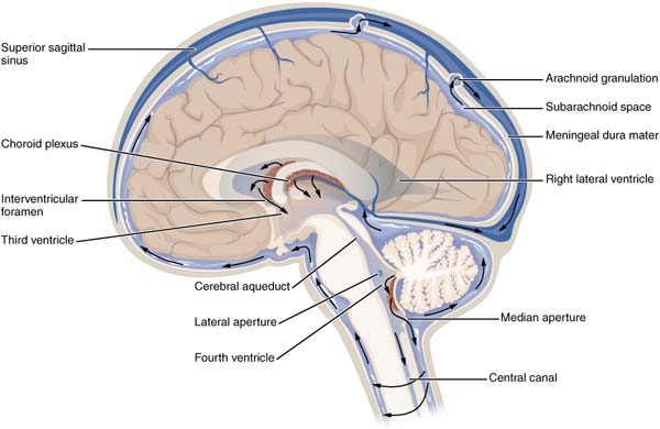
Figure 17.
View the Cerebrospinal Fluid Circulation video (opens in a new window) that shows the flow of CSF through the brain and spinal cord, and how it originates from the ventricles.
Disorders of the CNS
The supply of blood to the brain is crucial to its ability to perform many functions. Without a steady supply of oxygen, and to a lesser extent glucose, the nervous tissue in the brain cannot keep up its extensive electrical activity. These nutrients get into the brain through the blood, and if blood flow is interrupted, neurological function is compromised.
The common name for a disruption of blood supply to the brain is a stroke. It is caused by a blockage to an artery in the brain. The blockage is from some type of embolus: a blood clot, a fat embolus, or an air bubble. When the blood cannot travel through the artery, the surrounding tissue that is deprived starves and dies. Strokes will often result in the loss of very specific functions. A stroke in the lateral medulla, for example, can cause a loss in the ability to swallow.
Sometimes, seemingly unrelated functions will be lost because they are dependent on structures in the same region. Along with the swallowing in the previous example, a stroke in that region could affect sensory functions from the face or extremities because important white matter pathways also pass through the lateral medulla. Loss of blood flow to specific regions of the cortex can lead to the loss of specific higher functions, from the ability to recognize faces to the ability to move a particular region of the body. Severe or limited memory loss can be the result of a temporal lobe stroke.
Related to strokes are transient ischemic attacks (TIAs), which can also be called "mini-strokes." These are events in which a physical blockage may be temporary, cutting off the blood supply and oxygen to a region, but not to the extent that it causes cell death in that region. While the neurons in that area are recovering from the event, neurological function may be lost. Function can return if the area is able to recover from the event.
Glossary
- Amygdala
- nucleus deep in the temporal lobe of the cerebrum that is related to memory and emotional behavior
- Basal Forebrain
- nuclei of the cerebrum related to sensory stimuli and attention, loss of which is related to Alzheimer's disease
- Basal Nuclei
- nuclei of the cerebrum that are responsible for assessing cortical movement commands and comparing them with the general state of the individual through activity of dopamine neurons; largely related to motor functions, as seen in the symptoms of Parkinson's and Huntington's diseases
- Broca's Area
- region of the frontal lobe associated with the motor commands necessary for speech production and located only in the cerebral hemisphere responsible for language production
- Caudate
- nucleus deep in the cerebrum that is part of the basal nuclei
- Central Sulcus
- surface landmark of the cerebral cortex that marks the boundary between the frontal and parietal lobes
- Cerebral Cortex
- outer gray matter covering the forebrain, marked by wrinkles and folds known as gyri and sulci
- Cerebrum
- region of the adult brain that develops from the telencephalon and is responsible for higher neurological functions such as memory, emotion, and consciousness
- Cerebellum
- region of the adult brain connected primarily to the pons that developed from the metencephalon (along with the pons) and is largely responsible for comparing information from the cerebrum with sensory feedback from the periphery through the spinal cord
- Corpus Callosum
- large white matter structure that connects the right and left cerebral hemispheres
- Epithalamus
- region of the diecephalon containing the pineal gland
- Frontal Lobe
- region of the cerebral cortex directly beneath the frontal bone of the cranium
- Gyrus
- ridge formed by convolutions on the surface of the cerebrum or cerebellum
- Hippocampus
- gray matter deep in the temporal lobe that is very important for long-term memory formation
- Hypothalamus
- major region of the diencephalon that is responsible for coordinating autonomic and endocrine control of homeostasis
- Inferior Colliculus
- part of the brain stem auditory pathway
- Inferior Olive
- nucleus in the medulla that is involved in processing information related to motor control
- Lateral Sulcus
- surface landmark of the cerebral cortex that marks the boundary between the temporal lobe and the frontal and parietal lobes
- Limbic System
- structures at the edge (limit) of the boundary between the forebrain and hindbrain that are most associated with emotional behavior and memory formation
- Longitudinal Fissure
- large separation along the midline between the two cerebral hemispheres
- Occipital Lobe
- region of the cerebral cortex directly beneath the occipital bone of the cranium
- Parietal Lobe
- region of the cerebral cortex directly beneath the parietal bone of the cranium
- Postcentral Gyrus
- ridge just posterior to the central sulcus, in the parietal lobe, where somatosensory processing initially takes place in the cerebrum
- Precentral Gyrus
- primary motor cortex located in the frontal lobe of the cerebral cortex
- Prefrontal Lobe
- specific region of the frontal lobe anterior to the more specific motor function areas, which can be related to the early planning of movements and intentions to the point of being personality-type functions
- Premotor Area
- region of the frontal lobe responsible for planning movements that will be executed through the primary motor cortex
- Putamen
- nucleus deep in the cerebrum that is part of the basal nuclei
- Reticular Formation
- diffuse region of gray matter throughout the brain stem that regulates sleep, wakefulness, and states of consciousness
- Somatosensation
- general senses related to the body, usually thought of as the senses of touch, which would include pain, temperature, and proprioception
- Substantia Nigra Pars Compacta
- nuclei within the basal nuclei that release dopamine to modulate the function of the striatum; part of the motor pathway
- Sulcus
- groove formed by convolutions in the surface of the cerebral cortex
- Superior Colliculus
- responsible for aligning visual, auditory, and somatosensory spatial perceptions
- Tectum
- region of the midbrain, the roof of the cerebral aqueduct, which is subdivided into the inferior and superior colliculi
- Temporal Lobe
- region of the cerebral cortex directly beneath the temporal bone of the cranium
- Thalamus
- major region of the diencephalon that is responsible for relaying information between the cerebrum and the hindbrain, spinal cord, and periphery
Grant and Other Information

Except where otherwise noted, this work by The Community College Consortium for Bioscience Credentials is licensed under a Creative Commons Attribution 4.0 International License.
Text from BioBook licensed under CC BY NC SA and Boundless Biology Open Textbook licensed under CC BY SA.
Other text from OpenStaxCollege licensed under CC BY 3.0. Modified by Alice Rudolph, M.A. and Andrea Doub, M.S. for c3bc.
Instructional Design by Courtney A. Harrington, Ph.D., Helen Dollyhite, M.A. and Caroline Smith, M.A. for c3bc.
Media by Brittany Clark, Jose DeCastro, Jordan Campbell and Antonio Davis for c3bc.
This product was funded by a grant awarded by the U.S. Department of Labor's Employment and Training Administration. The product was created by the grantee and does not necessarily reflect the official position of the U.S. Department of Labor. The Department of Labor makes no guarantees, warranties, or assurances of any kind, express or implied, with respect to such information, including any information on linked sites and including, but not limited to, accuracy of the information or its completeness, timeliness, usefulness, adequacy, continued availability, or ownership.