Neurophysiology- The Action Potential
Anatomy & Physiology I
Neurophsiology- The Action Potential
Objectives
By the end of this section, the student will be able to:
- Describe the components of the membrane that establish the resting membrane potential.
- Describe the changes that occur to the membrane that result in the graded potential.
- Describe the changes that occur to the membrane that result in the action potential.
- Describe variations in the propagation of the action potential along the neuron.
Neurons even work sort of like electrical wires. Electrical wires conduct a flow of electrons we know as electricity or electrical current. Neurons conduct impulses that are electrical in nature. A neuron's impulse or signal is called an action potential. This action potential is electric in nature but it involves ion movement and not electron movement.
The term potential (membrane potential) is short for potential difference. If you have had a physics course and studied electricity, then you may know something about this topic already. Potential difference refers to a separation of charge. For example, in a battery there is a positive pole and a negative pole, these different areas represent separation of charge. An electric potential also exists across cell membranes. In muscle cells and neurons, there is a much greater potential difference than in other cells. There are more positive ions outside the neuron and muscle cell membrane than inside the cell. This is the separation of charge. The outside of the cell is more positive than the inside of the cell. We could say the inside of the cell is less positive but it is easier to refer to the inside of the cell as negative. Remember to think in terms of positivity, however.
The separation of charge is due to the membrane's differential permeability to sodium and potassium ions. The neuron and muscle cell membrane is very permeable to potassium ions and is relatively impermeable to sodium ions. Because of this, potassium ions are able to move back and forth across the plasma membrane along their diffusion gradient. However, sodium ions are not able to move back and forth across the membrane at will. Therefore, the sodium ions must remain outside the cell. This means that of the two ion species, there are both sodium and potassium ions outside the cell and mostly potassium ions inside the cell. This differential permeability of the membrane to these two ions creates the membrane potential.
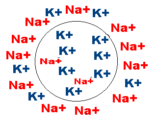
Figure 1. Electrically Active Cell Membranes. Cells in our bodies have more sodium (Na+) on the outside of the cells and more potassium (K+) on the inside of the cells. Since these are ions (charged atoms) they need a protein channel to cross the membrane, and are unable to move across on their own. Therefore, more Na+ stays on the outside and more K+ stays on the inside unless a chemical or electrical change occurs at the membrane.
This membrane potential can be measured using a device similar to a battery meter. However, instead of measuring the potential in volts, millivolts are used. A millivolt is 1/1000 a volt. To measure the membrane potential, an electrode must be inserted into the cell and just outside the cell membrane. This is a tricky procedure that requires a good microscope, a micromanipulator, microelectrodes, and a millivoltmeter. The potential that exists across a typical membrane has a magnitude of 70 mV. Because the inside of the cell is less positive than the outside, the membrane potential is said to be -70mV. The symbol in front of the number indicates the relative charge on the inside of the cell. The inside of the cell is 70 mV less positive than the outside of the cell. This is a typical resting membrane potential for a neuron. The resting membrane potential describes the potential of a cell membrane that is not being stimulated.
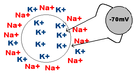
Figure 2. The nervous system can be divided into regions that are responsible for sensation (sensory functions) and for the response (motor functions). Sensory input needs to be integrated with other sensations. Some regions of the nervous system are termed integration or association areas; these use the interneurons of the central nervous system to communicate. The process of integration combines sensory perceptions and higher cognitive functions such as memories, learning, and emotion to produce a response.
The differentially (or semi-) permeable membrane creates the membrane potential. However, the membrane is not totally impermeable to sodium ions. Periodically, some sodium ions leak across the membrane and into the cell. Remember from the Cell membrane Module that the phospholipid bilayer contains protein channels to help in the transport of ions and large molecules across the membrane. This is one way the membrane in semi-permeable. These transmembrane channels consist of leak channels (like pores), chemically gated channels, voltage gated channels, etc. When we say that sodium leaks through the membrane, this happens through the protein leak channels. When an ion can move across the membrane due to electrical changes, this is due to the opening of voltage-gated channels.
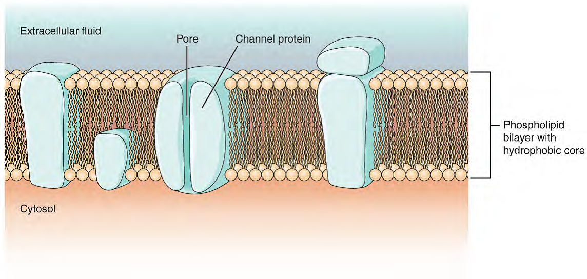
Figure 3. Cell Membrane and Transmembrane Proteins .The cell membrane is composed of a phospholipid bilayer and has many transmembrane proteins, including different types of channel proteins that serve as ion channels.
There are two forces acting on the sodium ions to move them into the cell, the force of diffusion and the electrical force. There is a high concentration of sodium outside the cell and a low concentration of sodium inside the cell. This concentration gradient would tend to move sodium ions into the cell. There is also an electrical gradient at work. The positively charged ions outside the cell repel one another. There are more positive ions outside the cell than inside the cell. The sodium ions tend to want to move into the cell along this electrical gradient. Although the membrane is relatively impermeable to sodium ions, they do tend to slip across the membrane periodically along their diffusion and electrical gradients. As these positive ions move into the cell, the membrane potential would become more and more positive if something didn't correct the problem. The resting membrane potential however is maintained at -70mV even though these positive ions keep slipping into the cell. This is because there is a sodium-potassium pump at work within the cell membrane. This pump actively and continually moves sodium ions out of the cell and potassium ions into the cell. For every three sodium ions it pumps out, it pumps in two potassium ions. This sodium-potassium pump is able to maintain the resting membrane potential at -70mV even though the sodium ions leak into the cells occasionally.
When the cell is not being stimulated, it is said to be resting. However, the resting membrane potential can change as a result of stimulation. There are two types of changes in membrane potential, action potentials and graded potentials. Action potentials are rapid changes in membrane potential that involve the entire cell. The action potential is the nerve impulse. Graded potentials are small changes in membrane potential. Graded potentials may lead to action potentials or they may inhibit action potentials. Let's look at action potentials first.
The Action Potential
Action Potentials are big changes in membrane potential. Remember in a resting cell, there are more positive ions outside the cell and fewer positive ions inside the cell. During an action potential, this situation is reversed. More positive ions are found inside the cell than outside the cell during an action potential. The inside of the cell actually becomes positive with respect to the outside. The membrane is said to depolarize. The membrane then repolarizes and returns to normal after a short period of hyperpolarization. Action potentials work according to the all-or-none principle. This means either an action potential happens or it doesn't happen, there is no in between. It's sort of like being pregnant. One is either pregnant or not pregnant, there is no in between. Or think about firing a gun, it either fires or it doesn't fire. It is not possible to barely shoot a bullet from the barrel of a gun. It comes out with complete force or not at all. Like firing a gun, action potentials have a threshold. To fire a gun, the trigger must be pulled. Triggers on some guns can be pulled back a little bit without the gun firing. However, once the trigger is pulled to a certain point, the hammer falls and the bullet fires. With action potentials, there is a threshold level that must be reached before the action potential will fire.
This means the magnitude of the stimulation on the cell must be large enough to create and action potential. If the stimulus strength is not at or above the threshold level, an action potential will not be fired. The threshold level for many neurons is -55mV. This means that the membrane must depolarize to -55mV or an action potential will not be fired. A stimulus may be strong enough to cause the membrane to depolarize to the threshold level or it may not be strong enough. In order for an action potential to fire, threshold must be reached.
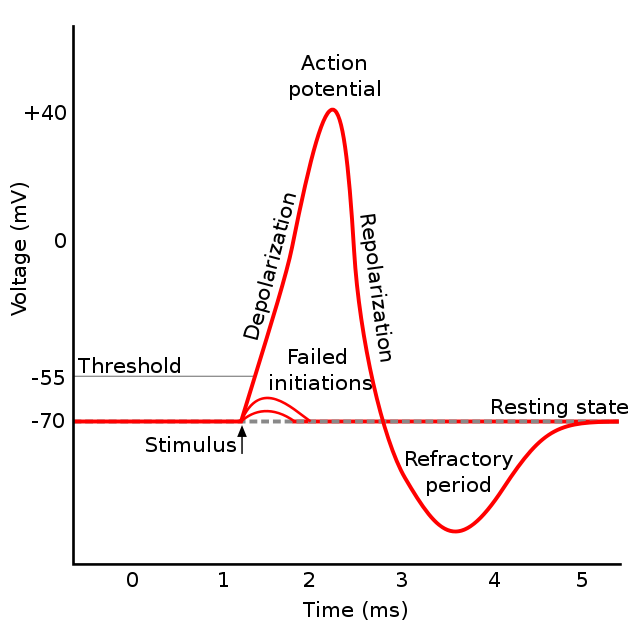
Figure 4. Graph of Action Potential. Plotting voltage measured across the cell membrane against time, the action potential begins with depolarization, followed by repolarization, which goes past the resting potential into hyperpolarization, and finally the membrane returns to rest.
The above figure shows how the membrane potential might change over time. The membrane was stimulated two times, but not to the threshold level. Therefore, an action potential was not fired. However, the third time the membrane was stimulated; the membrane did reach its threshold level. The result was an action potential. The action potential has three phases; the depolarization phase, the repolarization phase, and the after-hyperpolarization phase. As you can see, the action potential is a rapid change in membrane potential. In a matter of milliseconds, the membrane potential went from -70mV up to +30mV. The inside of the cell actually became more positive than the outside of the cell.
The threshold level may at first seem like some magical level at which the neuron fires an action potential. However, there is nothing magical too it at all. The threshold level is simply the point at which a very important positive feedback loop kicks in. Remember, positive feedback loops lead to the extreme. The cell experiences a change in one direction and responds by promoting a change in the same direction. Once the threshold level is reached (enough sodium ions were allowed in from the ligand-gated channels opening that the membrane potential gets up to -55mV), special voltage-gated sodium gates in the cell membrane open up. When these gates open, sodium ions stream into the cell along their electrical and diffusion gradients (because there was less inside than outside the cell). As sodium ions move into the cell, the inside of the cell becomes more positive (it depolarizes). This depolarization of the membrane causes even more sodium gates to open. When these additional sodium gates open, more sodium ions move into the cell. As more sodium ions move into the cell, the membrane depolarized even more. This additional depolarization causes even more sodium gates to open which causes more sodium ions to enter the cell which causes the cell membrane to depolarize even more... .... The net effect is the membrane becomes very permeable to sodium ions very quickly and sodium ions move into the cell along their diffusion and electrical gradients. This explains the depolarization phase.
Once the membrane reaches +30 mV, the voltage-gated sodium gates close and special voltage-gated potassium gates open. Sodium ions can no longer move into the cell and the sodium-potassium pump is continually pumping them out. The opening of the voltage-gated potassium channels causes the membrane to become even more permeable to potassium ions. As a result, more potassium ions leave the cell along their diffusion gradient (and electrical gradient at first). This movement of potassium ions out of the cell and the action of the sodium-potassium pump causes the cell membrane to repolarize.
The special potassium gates actually stay open for a few milliseconds after the membrane potential returns to resting. This causes the potassium ions to continue to diffuse out of the cell. The movement of potassium ions out of the cell results in the membrane potential becoming more negative than the resting membrane potential, which is called the after-hyperpolarization phase.
During the after-hyperpolarization phase, the cell is not responsive to stimuli of threshold strength. Because the membrane is hyperpolarized, it would require a stimulus of greater that threshold strength to fire another action potential. This is called the relative refractory period. During the relative refractory period, the cell is unresponsive to threshold strength stimuli. However, it will respond to suprathreshold strength stimuli.
There is also an absolute refractory period. During the absolute refractory period, the cell is completely unresponsive to successive stimuli, no matter how strong. The absolute refractory period corresponds with the depolarization and repolarization phases of the action potential. This is simply common sense; the cell can not fire two action potentials at once any more than a rifle with a single barrel can fire two bullets at exactly the same time.
The action potential is created at some point on the neuron's membrane. This action potential at this local area of the membrane triggers action potentials in the adjacent areas of the membrane. By this mechanism, the neurons entire membrane depolarizes and repolarizes. This process spreads across the membrane within milliseconds, even in a neuron with axons or dendrites over a meter long. Action potentials involve the entire cell. The entire cell depolarizes to +30mV and repolarizes to -70mV. Also, remember that action potentials are all-or-none, so once threshold is reached there is no stopping the signal from reaching the axon terminals.
The Graded Potential
Graded potentials are very different from action potentials. The term graded gives some insight to this fact. "Graded " describes the fact that the magnitude of the change in membrane potential is directly proportional to the magnitude of the stimulus. With graded potentials, a very weak stimulus will cause a very weak change in membrane potential. A very, very weak stimulus will cause a very, very weak change in membrane potential. A very, very, very weak stimulus will cause a very, very, very weak change in membrane potential. This is illustrated in the figure below. Graded is essentially the opposite of all or none. This also means that graded potentials do not have a threshold. No matter how small the stimulus, an equally small change in membrane potential will result (only a few sodium channels are opening so a smaller amount of sodium is being allowed into the cell).
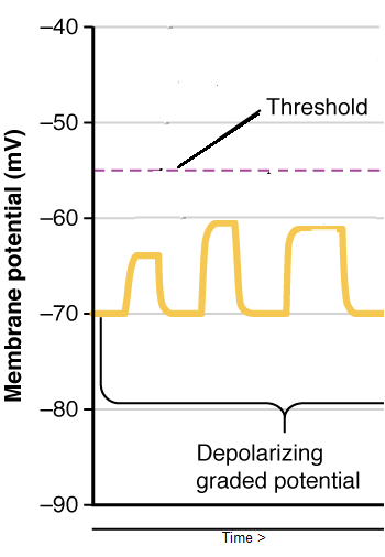
Figure 5. Graded Potential. These are small changes in the membrane potential. Notice the threshold potential is the critical level to which a membrane potential must be depolarized to initiate an action potential. If threshold is not reached, these graded, or local, portentials will decrease again, called decremental conduction.
Graded membrane potentials are also called local potentials. They are called local potentials because only a local area of the cell experiences the change in membrane potential. The entire cell is not affected by the graded potential. The change in membrane potential is strongest at the sight of stimulation. The magnitude of the change in membrane potential decreases as the distance from the sight of stimulation increases or they fizzle out. This is called decremental conduction.
Think of a stadium wave - you know when people in the baseball stadium put up their hands and then the next section does the same and it goes around the whole stadium (mostly an American phenomenon). When a section of people in the stadium want to start a wave, often the people around them are not 'open' to it and the signal, like a graded potential signal, fizzles out. If, however, enough of a the signal idea gets around (enough sodium is allowed into the neuron cell body) the neighboring people (or sodium channels in this case) are 'open' to the idea, all of the next section of the stadium will raise their hands over their heads (and lower them) and we can see a wave going all the way around the stadium - it is an all-or-none signal - as long as enough graded potentials are available and threshold is reached the action potential "wave" will go all the way down the neuron.
Graded changes in membrane potential may be in either a depolarizing or hyperpolarizing direction. An excitatory stimulus would depolarize the postsynaptic membrane, called an excitatory postsynaptic potential or EPSP. The depolarization would be the result of sodium gates opening up and sodium ions diffusing into the cell. The change is said to be excitatory because it results in the membrane potential being closer to threshold. An inhibitory stimulus would have the opposite effect. An inhibitory stimulus would hyperpolarize the postsynaptic membrane called an inhibitory postsynaptic potential or IPSP. The hyperpolarization would be the result of special (ligand-gated) potassium gates opening and potassium ions diffusing out of the cell. This is said to be inhibitory because the membrane is now farther away from threshold and is less likely to be able to fire an action potential. Graded potentials may be in either direction. These inhibitory channels could also be chloride ion (Cl-) channels, since there is more Chloride outside the cell than inside the cell. Amine neurotransmitters like glycine can open chloride channels if an inhibitory signal is needed.
Drug Interaction
Several drugs will either mimic an excitatory NT or an inhibitory NT and will contain chemicals that mimic these neurotransmitters. List NTs for Exc and Inh.
Some Neurotransmitters for chemically controlled channels and their functions:
- Acetylcholine (at "cholinergic" synapse) (EPSPs)
- Norepinephrine (noradrenalin) (at "adrenergic synapse") (EPSPs)
- Serotonin (can be either)- mood, appetite, depression
- Dopamine (can be either)- Parkinson's; personality, thoughts
- Gamma aminobutyric acid (GABA) (IPSPs)- anxiety
- Glycine (IPSPs) - inhibitory in spinal cord, brain; may help sleep quality
- Others (including NO and CO) - peptides - thirst, drinking behavior
Graded potentials are temporary changes in the membrane voltage, the characteristics of which depend on the size of the stimulus. Some types of stimuli cause depolarization of the membrane, whereas others cause hyperpolarization. It depends on the specific ion channels that are activated in the cell membrane.
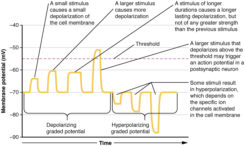
Figure 6. Graded Potentials. Graded potentials are temporary changes in the membrane voltage, the characteristics of which depend on the size of the stimulus. Some types of stimuli cause depolarization of the membrane, whereas others cause hyperpolarization. It depends on the specific ion channels that are activated in the cell membrane.
Graded potentials may also be summed. This means their effects may be combined. If two different stimuli stimulate the cell at about the same time and close by but in different places, their effects may be combined. This summation is called spatial summation. Think of a parent who has two children asking the same thing at the same time ("Dad, we want a popsicle. Daaadd"). Or, one stimulus may repeatedly stimulate a cell. This is called temporal summation - a single stimulation at one place on the neurolemma over and over. Think of when a single child repeatedly tugs at his Mother's sleeve saying "Mom, Mom, Mom, Mom,... " over and over - the parent could get to the point of saying "What?!" very loudly? She has reached her threshold, showing that the repeated stimulations may be added together to make an action potential, or in this case get a reaction. Whether or not a neuron fires an action potential or not depends on the combined effect of all incoming stimuli, both excitatory and inhibitory. Whether or not an action potential is fired depends on the combined effect of all incoming stimuli; if it is enough to reach the neuron's threshold level.

Figure 7. The excitation-contraction coupling effects of successive motor neuron signaling is added together which is referred to as wave summation.
Table 1. Contrasts ActionPotentials vs. Graded Potentials
| Action Potential |
Graded Potential |
|
All-or-none
|
Graded
|
|
Threshold
|
Graded
|
|
Involve the entire cell
|
Local
|
|
Refractory Periods
|
Can be summed
|
|
Always depolarization, repolarization, hyperpolarization
|
Can be in both directions
|
|
Entire cell experiences action potential
|
Decremental conduction
|
Propagation of the Action Potential Along the Neuron
The development of an action potential in one part of the neuron in turn creates action potentials in adjacent regions of the neurolemma going down the axon toward the terminals. The impulse spreads over the neurolemma like a wave (as mentioned before). One could picture this as a line of dominoes standing on end. If one domino is pushed the entire line of dominoes will fall, one after the next, going in one direction.
Video 1. View the YouTube video "Domino Analogy in a Neuron" (opens in new window)
The velocity of conduction down the neuron depends on the neuron's diameter and whether or not the neuron is myelinated (has a myelin sheath). Large diameter fibers conduct impulses faster than small diameter fibers. Think of a hallway with lots of students getting out of class at the same time. Which hallway width (or axon diameter) would allow faster movement of the crowd? The larger hallway does, as the larger axon width (diameter) allows for more movement of ions.
Cells that are myelinated exhibit very fast conduction due to a process called saltatory conduction. Saltatory conduction describes how the impulse 'jumps' from node of Ranvier to node of Ranvier. Saltatory means dancing or jumping, so I think of salsa dancing when I see this word and it makes me remember what this term refers to on the axon. The Schwann cells that wrap around the axons contain a substance called myelin. The myelin acts as an electrical insulator and therefore, the action potential must 'jump' these myelin sheaths to get from node to node.
Animation 1. View the YouTube animation "Saltatory Conduction" (opens in new window)
This is a much faster means of conduction and is also less energy expensive for the cell, because only the sodium channels that are at the small nodes have to be opened. Just as it is much faster to cross a room by taking giant steps instead of baby steps, the saltatory signal is faster and uses less energy. A signal that would have to open all of the sodium channels all the way down the axon (continuous conduction)would take a lot longer to get to the end. If one were to take baby steps to get across the room, instead of giant steps, it would take a lot longer to get there.
Video 2. View the YouTube video "Conduction Analogy" (opens in new window)
The conduction speed also depends on temperature - warmer body temperature allows for faster movement of action potentials versus a body that is in hypothermia.
Video 3. View the View the YouTube video "Characteristics of Life - Responsiveness" (opens in new window)

Glossary
- Absolute Refractory Period
- time during an action period when another action potential cannot be generated because the voltage-gated Na+ channel is inactivated
- All-or-None
- the principle that the response of a neuron or muscle fiber to a stimulus at any strength above the threshold is the same, that the muscle fiber or neuron cell responds completely or not at all
- Continuous Conduction
- slow propagation of an action potential along an unmyelinated axon owing to voltage-gated Na+ channels located along the entire length of the cell membrane
- Depolarization
- change in a cell membrane potential from rest toward zero
- Excitatory
- results in the membrane potential being closer to threshold
- Hyperpolarize
- a change in a cell's membrane potential that makes it more negative
- Inhibitory
- the membrane potentail gets farther away from threshold and is less likely to be able to send an action potential
- Leakage Channel
- ion channel that opens randomly and is not gated to a specific event, also known as a non-gated channel
- Ligand-gated Channels
- another name for an ionotropic receptor for which a neurotransmitter is the ligand
- Myelin Sheath
- lipid-rich layer of insulation that surrounds an axon, formed by oligodendrocytes in the CNS and Schwann cells in the PNS; facilitates the transmission of electrical signals
- Potential (membrane potential)
- distribution of charge across the cell membrane, based on the charges of ions
- Repolarization
- return of the membrane potential to its normally negative voltage at the end of the action potential
- Resting Membrane Potential
- the difference in voltage measured across a cell membrane under steady-state conditions, typically -70 mV
- Saltatory Conduction
- quick propagation of the action potential along a myelinated axon owing to voltage-gated Na+ channels being present only at the nodes of Ranvier
- Spatial Summation
- combination of graded potentials across the neuronal cell membrane caused by signals from separate presynaptic elements that add up to initiate an action potential
- Sodium Potassium Pump
- membrane-bound transporter found in nearly all cells that transports potassium ions into the cytoplasm from the extracellular fluid while simultaneously transporting sodium ions out of the cytoplasm to the extracellular fluid
- Temporal Summation
- combination of graded potentials at the same location on a neuron resulting in a strong signal from one input
- Threshold
- millivolt level that must be reached before the action potential can be produced
- Voltage-gated Channel
- ion channel that opens because of a change in the charge distributed across the membrane where it is located
Grant and Other Information

Except where otherwise noted, this work by The Community College Consortium for Bioscience Credentials is licensed under a Creative Commons Attribution 4.0 International License.
Text from Michael Ayers, M.S. for c3bc.
Other text from BioBook licensed under CC BY NC SA and Boundless Biology Open Textbook licensed under CC BY SA.
Other text from OpenStaxCollege licensed under CC BY 3.0. Modified by Alice Rudolph, M.A. and Andrea Doub, M.S. for c3bc.
Instructional Design by Courtney A. Harrington, Ph.D., Helen Dollyhite, M.A. and Caroline Smith, M.A. for c3bc.
Media by Brittany Clark, Jose DeCastro, Jordan Campbell and Antonio Davis for c3bc.
This product was funded by a grant awarded by the U.S. Department of Labor's Employment and Training Administration. The product was created by the grantee and does not necessarily reflect the official position of the U.S. Department of Labor. The Department of Labor makes no guarantees, warranties, or assurances of any kind, express or implied, with respect to such information, including any information on linked sites and including, but not limited to, accuracy of the information or its completeness, timeliness, usefulness, adequacy, continued availability, or ownership.
;