Gross and Microscopic Anatomy of Nerve Tissue- Neuron Anatomy and Physiology
Anatomy & Physiology I
Gross and Microscopic Anatomy of Nerve Tissue - Neuron Anatomy and Physiology
Objectives
By the end of the section the student will be able to:
- Describe the basic structure of a neuron
- Describe the functions of each part of the neuron
The cell involved with transmitting information in the nervous system is called a "neuron." There are three kinds of neurons functionally speaking. These are the sensory neurons, interneurons, and motor neurons. Sensory neurons are afferent neurons that conduct information into the CNS. Motor neurons are efferent neurons that conduct information out of the CNS. The interneurons are housed entirely within the CNS and are the ones responsible for integration of information.

Figure 1. Functions of the Nervous System. Sensory input from a receptor (monitors internal and external stimuli from the skin or organs) sends an impulse signal to the Central Nervous System and an Interneuron there where the brain and spinal cord will process sensory input and send out a response. The motor output will send a signal to control muscles and glands, which are the effectors.
The Neuron
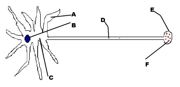
In the diagram below, several parts of the multipolar neuron are labeled. The dendrites (A) and the axon (D) are cytoplasmic extensions of the cell. Dendrites conduct impulses to the cell body (B). They are very long in sensory neurons (up to a meter or so) and much shorter in motor neurons. Axons conduct information away (think " Axon Away") from the cell body they are very short in sensory neurons and long in motor neurons (up to a meter or so). The soma or cell body contains the nucleus of the cell. Sensory neurons are unipolar and their cell bodies are found in structures bilateral to the spinal cord called dorsal root ganglia. Cell bodies of motor neurons are found in the anterior horn of the spinal cord. We will discuss spinal cord anatomy later. At the end of the axon is a structure called the synaptic end-bulb (E). Within the synaptic end-bulb are structures called synaptic vesicles (F). The axon terminal with synaptic vesicles contain chemicals called neurotransmitter substances. Axons and dendrites are analogous to "wires" because they transmit information and they run throughout our bodies much like you have wires that run within walls of your home. Axons and dendrites are found in bundles throughout our body. These bundles of axons and dendrites are called nerves (or referred to as 'tracts' in the CNS).

Figure 2. Labeled Multipolar Neuron. The dendrites (A) and the axon (D) are cytoplasmic extensions of the cell. Dendrites conduct impulses to the cell body (B). They are very long in sensory neurons (up to a meter or so) and much shorter in motor neurons. Axons conduct information away (think " Axon Away") from the cell body they are very short in sensory neurons and long in motor neurons (up to a meter or so). The soma or cell body contains the nucleus of the cell.

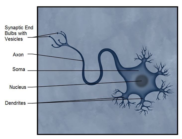
Figure 3. Another View of a Neuron showing the Synaptic End Bulbs with Vesicles, Axon, Soma, Nucleus and Dendrites

Figure 4. An actual micrograph of a neuron, showing the soma and nucleus.


Glossary
- Axon
- single stretch of the axon insulated by myelin and bounded by nodes of Ranvier at either end (except for the first, which is after the initial segment, and the last, which is followed by the axon terminal)
- Axon terminal with synaptic vesicles
- end of the axon, where there are usually several branches extending toward the target cell
- Dendrites
- cytoplasmic extension of the neuron receive signals and sends to the cell body
- Dorsal root ganglia
- sensory ganglion attached to the posterior nerve root of a spinal nerve
- Multipolar
- shape of a neuron that has multiple processes—the axon and two or more dendrites
- Nerves
- bundles of axons and dendrites found in the PNS throughout our body
- Synaptic end bulb
- swelling at the end of an axon where neurotransmitter molecules are released onto a target cell across a synapse
- Soma
- the cell body of the neuron
- Tracts
- bundles of axons and dendrites found in the CNS
- Unipolar
- shape of a neuron which has only one process that includes both the axon and dendrite
Grant and Other Information

Except where otherwise noted, this work by The Community College Consortium for Bioscience Credentials is licensed under a Creative Commons Attribution 4.0 International License.
Text from Micahel Ayers, M.S. for c3bc
Other text from BioBook licensed under CC BY NC SA and Boundless Biology Open Textbook licensed under CC BY SA.
Other text from OpenStaxCollege licensed under CC BY 3.0. Modified by Alice Rudolph, M.A. and Andrea Doub, M.S. for c3bc.
Instructional Design by Courtney A. Harrington, Ph.D., Helen Dollyhite, M.A. and Caroline Smith, M.A. for c3bc.
Media by Brittany Clark, Jose DeCastro, Jordan Campbell and Antonio Davis for c3bc.
This product was funded by a grant awarded by the U.S. Department of Labor's Employment and Training Administration. The product was created by the grantee and does not necessarily reflect the official position of the U.S. Department of Labor. The Department of Labor makes no guarantees, warranties, or assurances of any kind, express or implied, with respect to such information, including any information on linked sites and including, but not limited to, accuracy of the information or its completeness, timeliness, usefulness, adequacy, continued availability, or ownership.
;


