Motor Units, Skeletal Muscle Fibers and Effects of Exercise
Anatomy & Physiology I
Motor Units, Skeletal Muscle Fibers and Effects of Exercise
Objectives
- Students will be able to describe the structure and function of a motor unit, motor unit recruitment and the phases of twitch contraction.
- Students will be able to apply this information to explain how the frequency of stimulation affects muscle tension and leads to muscle tone, and isotonic and isometric contractions.
- Students will be able to compare the structure and function of the three types of skeletal muscle fibers (cells).
- Students will be able to describe the effects of exercise on different types of skeletal muscle fibers.
Motor Units
As you have learned, every skeletal muscle cell must be innervated by a somatic motor neuron in order to contract. This somatic motor neuron will branch and innervate several skeletal muscle cells; additionally, each skeletal muscle cell is innervated by only one somatic motor neuron. A single somatic motor neuron and all the skeletal muscle cells that it innervates are known as a motor unit. The size of a motor unit is variable depending on the nature of the muscle.
A small motor unit is an arrangement where a single motor neuron supplies a small number of muscle cells in a whole skeletal muscle. Small motor units permit very fine motor control of the muscle. The best example in humans is the small motor units of the extraocular eye muscles that move the eyes. There are thousands of muscle fibers in each muscle, but every six or so fibers are supplied by a single motor neuron, as the axons branch to form synaptic connections at their individual neuromuscular junctions. This allows for exquisite control of eye movements so that both eyes can quickly focus on the same object. Small motor units are also involved in the many fine movements of the fingers and thumb of the hand for grasping, texting, etc.
A large motor unit is an arrangement where a single motor neuron supplies a large number of muscle fibers in a muscle. Large motor units are concerned with simple, or "gross," movements, such as powerfully extending the leg at the knee joint. The best example is the large motor units of the thigh muscles or back muscles, where a single motor neuron will supply thousands of muscle fibers in a muscle, as its axon splits into thousands of branches.
There is a wide range of motor units within many skeletal muscles, which gives the nervous system a wide range of control over the muscle. The small motor units in the muscle will have smaller, more excitable motor neurons, firing first to their skeletal muscle fibers, which also tend to be the smallest. Activation of these smaller motor units, results in a relatively small degree of contractile strength (tension) generated in the muscle. As more strength is needed, larger motor units, with bigger, higher-threshold motor neurons are enlisted to activate larger muscle fibers. This increasing activation of motor units produces an increase in muscle contraction known as recruitment. As more motor units are recruited, the muscle contraction grows progressively stronger. In some muscles, the largest motor units may generate a contractile force of 50 times more than the smallest motor units in the muscle. This allows a coffee mug to be picked up using the biceps brachii arm muscle with minimal force, and a couch to be lifted by the same muscle by recruiting the largest motor units. Another way to think about recruitment is to remember back to a time when you have moved residences. When you move you pack everything into boxes and recruit help to aid you in moving which objects ... which needs more help: the box full of toiletries or the sectional couch? You recruit help for the couch because it requires more force to move that you alone can produce. If each person helping in the move, including yourself, are considered to be individual single motor units, it becomes clear only one motor unit is required to move smaller, lighter objects. But when moving very large, heavy objects more motor units are required, producing a great deal of force and successfully moving the object.
Recruitment
Each motor unit contracts in an all or none fashion - meaning each cell stimulated by the neuron contracts fully or not at all. If the load you are moving is light, then few motor units will be stimulated to produce the right amount of tension while fully contracting. When necessary, the maximal number of motor units in a muscle can be recruited simultaneously, producing the maximum force of contraction for that muscle, but this cannot last for very long because of the energy requirements to sustain the contraction. To prevent complete muscle fatigue, motor units are generally not all simultaneously active, but instead some motor units rest while others are active, which allows for longer muscle contractions. The nervous system uses recruitment as a mechanism to efficiently utilize a skeletal muscle.
To clarify, skeletal muscles are rarely completely relaxed, or flaccid. Even if a muscle is not producing movement, it is contracted a small amount to maintain its contractile proteins and produce muscle tone. The tension produced by muscle tone allows muscles to continually stabilize joints and maintain posture. Muscle tone is accomplished by a complex interaction between the nervous system and skeletal muscles that results in the activation of a few motor units at a time, most likely in a cyclical manner. In this manner, muscles never fatigue completely, as some motor units can recover while others are active.
Muscle Twitch and Summation (Myograms)
A single action potential from a somatic motor neuron will produce a single contraction in the muscle fibers of its motor unit. As we have stated before, this isolated singular contraction/relaxation cycle is called a twitch. A twitch can last for a few milliseconds or 100 milliseconds, depending on the muscle type. The tension produced by a single twitch can be measured by a myogram, an instrument that measures the amount of tension produced over time. Each twitch undergoes three phases. The first phase is the latent period, during which the action potential is being transmitted along the sarcolemma and Ca2+ ions are released from the SR (Steps # 7 - 11). This is the phase during which excitation and contraction are being coupled but contraction has yet to occur. The contraction phase occurs next. The Ca2+ ions in the sarcoplasm have bound to troponin, tropomyosin has shifted away from myosin-binding sites, cross-bridges formed, and sarcomeres are actively shortening to the point of peak tension (Steps # 12 - 14). The last phase is the relaxation phase, when tension decreases as contraction stops. Ca2+ ions are actively pumped out of the sarcoplasm into the sarcoplasmic reticulum, and cross-bridge cycling stops, returning the muscle fibers to their resting state.
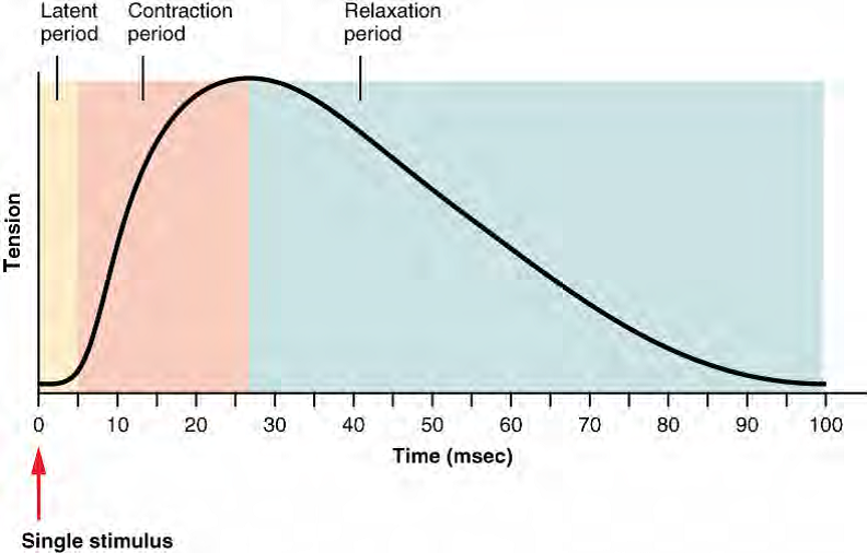
Figure 1: This Myogram of a Muscle Twitch shows a graph of an increasing line to a peak during the contraction period then a slower decline afterwards in the Relaxation period. A single muscle twitch has a latent period, a contraction phase when tension increases, and a relaxation phase when tension decreases. During the latent period (short flat line at the beginning of the graph), the action potential is being spread along the sarcolemma. During the contraction phase (a rapid increase in the slope of the graph), Ca++ ions in the sarcoplasm cause the steps of contraction to occur. During the relaxation phase, tension decreases (the slope on the graph goes down) as Ca++ ions are pumped out of the sarcoplasm and cross-bridges detach as contraction stops.
Although a person can experience a muscle "twitch," a single twitch does not producesignificant muscle tension in a living body. Meaning that if I wanted to lift my coffee mug, there is not enough tension produced by single muscle twitch to support this motion. A series of action potentials to the muscle fibers is necessary to produce a muscle contraction that can produce work such as lifting the mug. Normal muscle contraction is more sustained, and it can be modified by input from the nervous system to produce varying amounts of force; this is called a graded muscle response. The frequency of action potentials from a somatic motor neuron and the number of somatic motor neurons transmitting action potentials both affect the tension produced in skeletal muscle.
The rate at which a motor neuron fires action potentials affects the tension produced in the skeletal muscle. If the fibers are stimulated while a previous twitch is still occurring, the second twitch will be stronger. This response is called wave summation, because the excitation-contraction coupling effects of successive motor neuron signaling is summed, or added together. At the molecular level, summation occurs because the second stimulus triggers the release of more Ca2+ ions, which become available to activate additional sarcomeres while the muscle is still contracting from the first stimulus. Summation results in greater contraction of the motor unit. More simply - think back to your Steps # 1-14: the more action potentials that travel down the somatic motor neuron, the more ACh released into the synaptic cleft => the more action potentials that travel down the sarcolemma => the more calcium released from the sarcoplasmic reticulum = > more contraction => more force production due to wave summation.

Figure 2. Wave Summation Myogram. The excitation-contraction coupling effects of successive motor neuron signaling is added together which is referred to as wave summation. The bottom of each wave, the end of the relaxation phase, represents the point of stimulus and will start a new signal before the first one completely relaxed.
If the frequency of somatic motor neuron signaling increases, summation and subsequent muscle tension in the motor unit continues to rise until it reaches a peak point. The tension at this point is about three to four times greater than the tension of a single twitch, a state referred to as incomplete or unfused tetanus. During incomplete tetanus, the muscle goes through quick cycles of contraction with a short relaxation phase for each. Notice that force production increases but then declines slightly before beginning to increase again. This decline in force production is the partial relaxation between contractions and lends a sawtooth appearance to incomplete tetanus on the myogram. If the stimulus frequency is so high that the relaxation phase disappears completely, contractions become continuous in a process called complete tetanus. During tetanus, the concentration of Ca2+ ions in the sarcoplasm allows virtually all of the sarcomeres to form cross-bridges and shorten, so that a contraction can continue uninterrupted (until the muscle fatigues and can no longer produce tension). A myogram of complete tetanus shows a steep rise in tension production that plateaus when the muscle is contracting fully, and then a decline in force production that is caused when the muscle begins to fatigue from the continuous contraction.
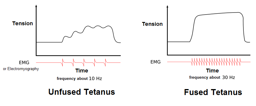
Figure 3. Myograms for Unfused Tetanus versus Fused Tetanus. During incomplete tetanus, the muscle goes through quick cycles of contraction with a short (incomplete) relaxation phase for each. If the stimulus frequency is so high that there is no relaxation phase (it disappears completely), contractions become continuous in a process called complete tetanus.
Isotonic and Isometric contractions
To move an object, referred to as load, the sarcomeres in the muscle fibers of the skeletal muscle must shorten. The force generated by the contraction of the muscle (or shortening of the sarcomeres) is called muscle tension. However, muscle tension also is generated when the muscle is contracting against a load that does not move, resulting in two main types of skeletal muscle contractions: isotonic contractions and isometric contractions.
In isotonic contractions, as the length of the muscle changes and tension is produced a load is moved. There are two types of isotonic contractions: concentric and eccentric. A concentric contraction involves the muscle shortening to move a load. Think concentric circles get smaller and smaller as you move toward the center, so a concentric contraction the sarcomeres get shorter and shorter. An example of this is the biceps brachii muscle contracting when a hand weight is brought upward with increasing muscle tension. As the biceps brachii contract, the angle of the elbow joint decreases as the forearm is brought toward the body. Here, the biceps brachii contracts as sarcomeres in its muscle fibers are shortening and cross-bridges form. You can feel the shortening happen. Place your right hand on your left biceps brachii as the left arm is fully extended. Now flex your arm, keeping your right hand in place. You can feel the muscle get shorter and begin to bunch up under your hand. This is the concentric isotonic contraction. An eccentric contraction occurs as the muscle tension diminishes and the muscle lengthens. In this case, the hand weight is lowered in a slow and controlled manner as tension is released from the biceps brachii, the angle of the elbow joint increases. Eccentric contractions are also used for movement and balance of the body. Just like before, you can feel the elongation happen. Place your right hand on your left biceps brachii as the left arm is flexed. Now extend your arm, keeping your right hand in place. You can feel the muscle get longer and begin to relax under your hand while still maintaining enough force to hold the weight. This is the eccentric isotonic contraction. Both of these movements involve the muscle changing length and a load being moved as a result.
An isometric contraction occurs as the muscle produces tension without changing the angle of a skeletal joint. Isometric contractions involve sarcomere shortening and increasing muscle tension, but do not move a load, as the force produced cannot overcome the resistance provided by the load. For example, if one attempts to lift a hand weight that is too heavy, there will be sarcomere activation and shortening to a point, and ever-increasing muscle tension, but no change in the angle of the elbow joint. In everyday living, isometric contractions are active in maintaining posture and maintaining bone and joint stability. However, holding your head in an upright position occurs not because the muscles cannot move the head, but because the goal is to remain stationary and not produce movement. This is the same principle behind the idea of the yoga workout; muscles produce tension and are being exercised but the goal is to remain stationary and resist movement. Most actions of the body are the result of a combination of isotonic and isometric contractions working together to produce a wide range of outcomes.
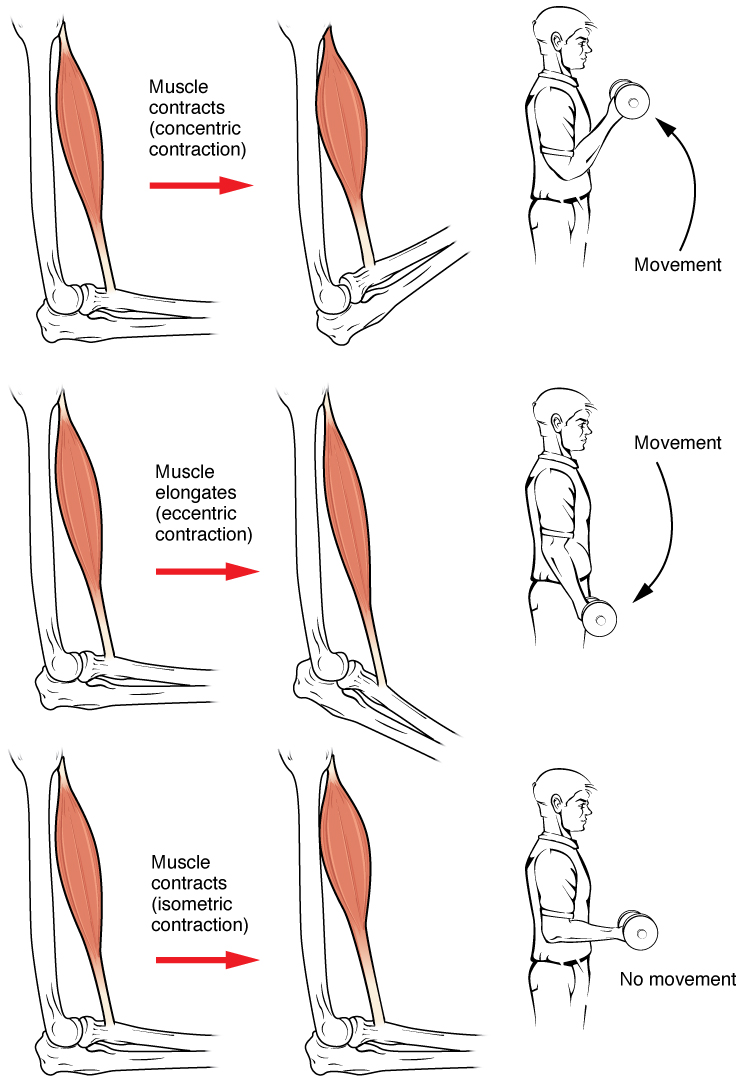
Figure 4. During isotonic contractions, muscle length changes to move a load. The concentric contraction shortens the muscle to move the load. The eccentric contraction lengthens the muscle to move the load, while maintaining enough force to hold the load. During isometric contractions, muscle length does not change because the load exceeds the tension the muscle can generate.
Types of Muscle Fibers
Imagine that it is Thanksgiving and as you are passing around all of the delicious calorie filled food you are asked the ultimate question ... Would you like white or dark meat? The choice is obvious but have you ever wondered why there is a difference between the two and what is responsible for that difference. Animals have specific patterns of movement characteristic to a particular species. Dark meat is composed of muscle cells that are built for endurance type activities; white meat contains cells built for short powerful movements. Let's think about the turkey. Turkeys and chickens primarily walk/run everywhere they go. While these birds can fly they can only sustain that type of activity for short periods of time. Based on this behavioral pattern, as anatomists we can infer that we will see cells built for endurance in the muscles that are used for prolonged periods of time; these cells compose the dark meat in legs and thighs of these birds. We can also infer that since flight is an activity that cannot be sustained for long periods of time, we will see cells built for quick bursts of energy in the muscle that cause flight; these cells compose the white meat in the breast of these birds. Now, think about a bird of flight like ducks or geese. What kind of meat would you find in these birds?
Now that we have solved that question it brings up another very interesting one, if animals' muscles are classified as dark and white meat, as humans do we have the same classification. We are all animals after all. But unlike the animals we just discussed, humans do not have specific behavioral patterns. You can use one muscle for a variety of actions - you can pick up a feather, then a couch, then hold a book out to someone, then drive, etc. This is why whole skeletal muscles like the biceps brachii are not made up of only one type of skeletal muscle cell, there are in fact 3 different types of skeletal muscle cells that are present in one whole skeletal muscle. There are two criteria to consider when classifying the types of skeletal muscle cells: how fast some cells contract relative to others and how cells produce ATP. The speed of contraction is dependent on how quickly myosin's ATPase hydrolyzes ATP to produce cross-bridges. Fast fibers hydrolyze ATP approximately twice as quickly as slow fibers, resulting in much quicker cross-bridge cycling (which pulls the thin filaments toward the center of the sarcomeres at a faster rate). How cells produce ATP is dependent on the primary metabolic pathway that the cell uses and will determine whether the cell is classified as oxidative or glycolytic. If a cell primarily produces ATP through aerobic (with oxygen) pathways it is oxidative. More ATP can be produced during each metabolic cycle, making the cell more resistant to fatigue. Oxygen debt is the amount of oxygen needed to compensate for ATP produced without oxygen during muscle contraction. The oxidative fibers contain many more mitochondria than the glycolytic fibers, because aerobic metabolism occurs in the mitochondria. Glycolytic fibers primarily create ATP through anaerobic (without oxygen) glycolysis, which produces less ATP per cycle. As a result, glycolytic fibers fatigue at a quicker rate. Using these criteria, the three main types of skeletal muscle cells are: slow oxidative, fast oxidative - glycolytic, and fast glycolytic.
Slow oxidative (SO) fibers contract relatively slowly and use aerobic respiration to produce ATP. The SO fibers possess a large number of mitochondria and are capable of contracting for longer periods because of the large amount of ATP they can produce, but they have a relatively small diameter and do not produce a large amount of tension. SO fibers are extensively supplied with blood capillaries to supply O2 from the red blood cells in the bloodstream. The SO fibers also possess myoglobin, an O2-carrying molecule similar to O2-carrying hemoglobin in the red blood cells. The myoglobin stores some of the needed O2 within the fibers themselves (and gives SO fibers their red color). All of these features allow SO fibers to produce large quantities of ATP, which can sustain muscle activity without fatiguing for long periods of time. The fact that SO fibers can function for long periods without fatiguing makes them useful in maintaining posture, producing isometric contractions, stabilizing bones and joints, and making small movements that happen often but do not require large amounts of energy.
Fast oxidative - glycolytic (FOG) fibers have fast contractions and primarily use aerobic respiration, but because they may switch to anaerobic respiration (glycolysis), can fatigue more quickly than SO fibers. FOG fibers are sometimes called intermediate fibers because they possess characteristics that are intermediate between fast fibers and slow fibers. They produce ATP relatively quickly, more quickly than SO fibers, and thus can produce relatively high amounts of tension. They are oxidative because they produce ATP aerobically, possess high amounts of mitochondria, and do not fatigue quickly. However, FO fibers do not possess significant myoglobin, giving them a lighter color than the red SO fibers. FO fibers are used primarily for movements, such as walking, that require more energy than postural control but less energy than an explosive movement, such as sprinting. FO fibers are useful for this type of movement because they produce more tension than SO fibers but they are more fatigue-resistant than FG fibers.
Lastly, fast glycolytic (FG) fibers have fast contractions and primarily use anaerobic glycolysis. The FG fibers fatigue more quickly than the others. FG fibers primarily use anaerobic glycolysis as their ATP source. They have a large diameter and possess high amounts of glycogen, which is used in glycolysis to generate ATP quickly to produce high levels of tension. Because they do not primarily use aerobic metabolism, they do not possess substantial numbers of mitochondria or significant amounts of myoglobin and therefore have a white color. FG fibers are used to produce rapid, forceful contractions to make quick, powerful movements. These fibers fatigue quickly, permitting them to only be used for short periods.
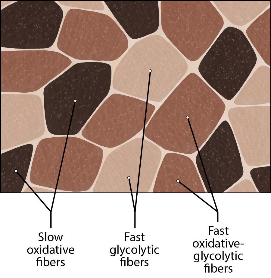
Figure 5. Types of Muscle Fibers. Please note that Slow Oxidative Fibers are the darkest in color, while Fast Oxidative Glycolytic Fibers are lighter and Fast Glycolytic Fibers are the lightest. Are you able to explain this, based on what you've learned?
As we stated earlier most skeletal muscles in a human possess a mixture of each cell type, although in varying proportions. The predominant fiber type in a muscle is determined by the primary function of the muscle. This is why you can train a particular muscle for a specific activity. If you plan to run a marathon, you must train your muscles to be more fatigue resistant by running a short distance and gradually increasing that distance as the weeks pass by. This training converts FOG and FG cell types in a skeletal muscle into SO cells. In doing this the whole muscle will be more fatigue resistant as a whole and you can then run the marathon. If you stop running the need for all of these SO cells decreases and the extra SO cells will be converted back into FOG and FG cell types.


Effects of Exercise on Skeletal Muscle Fibers
You may have heard the saying, "if you don't use it - you lose it," this certainly holds true when it comes to muscle size and strength. Physical training alters the appearance of skeletal muscles and can increase muscle performance. Conversely, a lack of use can result in decreased performance and muscle appearance. A whole skeletal muscle changes appearance because the muscle cells that compose that particular skeletal muscle can change in size. The cell diameter increases due to structural proteins being added to muscle cells in a process called hypertrophy. I must clarify that structural proteins are being added to existing cells; new cells are not formed when muscles grow. The number of skeletal muscle fibers in a given muscle is genetically determined and does not change. D ecreased use of a skeletal muscle results in atrophy, where the number of sarcomeres and structural proteins are lost (but not the number of muscle cells) decreasing muscle mass. It is common for a limb in a cast to show atrophied muscles when the cast is removed, and certain diseases, such as polio, show atrophied muscles.
Endurance Exercise
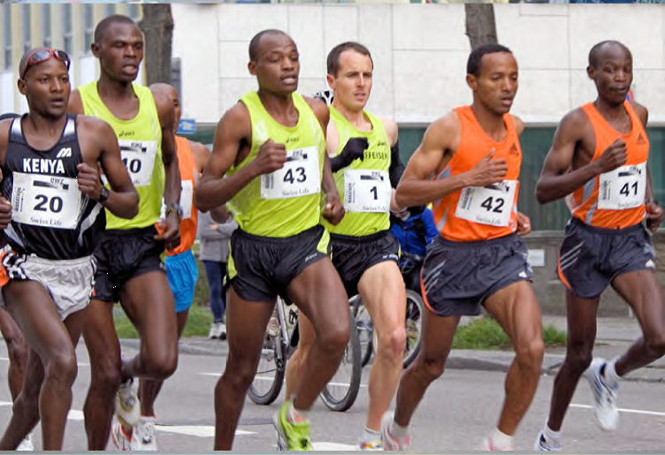
Figure 6. Long-distance runners have a large number of SO fibers and relatively few FO and FG fibers.
Slow fibers are predominantly used in endurance exercises that require little force but involve numerous repetitions. The aerobic metabolism used by slow-twitch fibers allows them to maintain contractions over long periods. Endurance training modifies these slow fibers to make them even more efficient by producing more mitochondria to enable more aerobic metabolism and more ATP production. Endurance exercise can also increase the amount of myoglobin in a cell, as increased aerobic respiration increases the need for oxygen. Myoglobin is found in the sarcoplasm and acts as an oxygen storage supply for the mitochondria.
The training can trigger the formation of more extensive capillary networks around the fiber, a process called angiogenesis, to supply oxygen and remove metabolic waste. To allow these capillary networks to supply the deep portions of the muscle, muscle mass does not greatly increase in order to maintain a smaller area for the diffusion of nutrients and gases. All of these cellular changes result in the ability to sustain low levels of muscle contractions for greater periods without fatiguing.
The proportion of SO muscle fibers in muscle determines the suitability of that muscle for endurance, and may benefit those participating in endurance activities. Postural muscles have a large number of SO fibers and relatively few FO and FG fibers, to keep the back straight. Endurance athletes, like marathon-runners also would benefit from a larger proportion of SO fibers, but it is unclear if the most-successful marathoners are those with naturally high numbers of SO fibers, or whether the most successful marathon runners develop high numbers of SO fibers with repetitive training. Endurance training can result in overuse injuries such as stress fractures and joint and tendon inflammation.
Resistance Exercise

Figure 7. Body builders have a large number of FG fibers and relatively few FO and SO fibers.
Resistance exercises, as opposed to endurance exercise, require large amounts of FG fibers to produce short, powerful movements that are not repeated over long periods. The high rates of ATP hydrolysis and cross-bridge formation in FG fibers result in powerful muscle contractions. Muscles used for power have a higher ratio of FG to SO/FO fibers, and trained athletes possess even higher levels of FG fibers in their muscles. Resistance exercise affects muscles by increasing the formation of myofibrils, thereby increasing the thickness of muscle fibers. This added structure causes hypertrophy, or the enlargement of muscles, exemplified by the large skeletal muscles seen in body builders and other athletes. Because this muscular enlargement is achieved by the addition of structural proteins, athletes trying to build muscle mass often ingest large amounts of protein.
Except for the hypertrophy that follows an increase in the number of sarcomeres and myofibrils in a skeletal muscle, the cellular changes observed during endurance training do not usually occur with resistance training. There is usually no significant increase in mitochondria or capillary density. However, resistance training does increase the development of connective tissue, which adds to the overall mass of the muscle and helps to contain muscles as they produce increasingly powerful contractions. Tendons also become stronger to prevent tendon damage, as the force produced by muscles is transferred to tendons that attach the muscle to bone.
For effective strength training, the intensity of the exercise must continually be increased. For instance, continued weight lifting without increasing the weight of the load does not increase muscle size. To produce ever-greater results, the weights lifted must become increasingly heavier, making it more difficult for muscles to move the load. The muscle then adapts to this heavier load, and an even heavier load must be used if even greater muscle mass is desired.
If done improperly, resistance training can lead to overuse injuries of the muscle, tendon, or bone. These injuries can occur if the load is too heavy or if the muscles are not given sufficient time between workouts to recover or if joints are not aligned properly during the exercises. Cellular damage to muscle fibers that occurs after intense exercise includes damage to the sarcolemma and myofibrils. This muscle damage contributes to the feeling of soreness after strenuous exercise, but muscles gain mass as this damage is repaired, and additional structural proteins are added to replace the damaged ones. Overworking skeletal muscles can also lead to tendon damage and even skeletal damage if the load is too great for the muscles to bear.
Performance-Enhancing Substances
Some athletes attempt to boost their performance by using various agents that may enhance muscle performance. Anabolic steroids are one of the more widely known agents used to boost muscle mass and increase power output. They are taken by athletes and mimic the hormone, testosterone, which is a hormone that increases muscle size by stimulating the production of intracellular proteins stimulating muscle formation and leading to increased muscle mass. This increase in these proteins leads to an increase in the size and strength of the muscle. But they come with serious side effects. Anabolic steroid use has been linked to infertility, aggressive behavior, cardiovascular disease, brain cancer, and many other disorders.
Endurance athletes may also try to boost the availability of oxygen to muscles to increase aerobic respiration by using substances such as erythropoietin (EPO), a hormone normally produced in the kidneys, which triggers the production of red blood cells. The extra oxygen carried by these blood cells can then be used by muscles for aerobic respiration.
Human growth hormone (hGH) is another supplement, and although it can facilitate building muscle mass, its main role is to promote the healing of muscle and other tissues after strenuous exercise. Increased hGH may allow for faster recovery after muscle damage, reducing the rest required after exercise, and allowing for more sustained high-level performance.
Creatine phosphate is another supplement that increases power output by providing quick bursts of ATP to muscles in the initial stages of contraction. Increasing the amount of creatine available to cells is thought to produce more ATP and therefore increase explosive power output, although its effectiveness as a supplement has been questioned.
Although performance-enhancing substances often do improve performance, most are banned by governing bodies in sports and are illegal for nonmedical purposes. Their use to enhance performance raises ethical issues of cheating because they give users an unfair advantage over nonusers. A greater concern, however, is that their use carries serious health risks. The side effects of these substances are often significant, nonreversible, and in some cases fatal. The physiological strain caused by these substances is often greater than what the body can handle, leading to effects that are unpredictable and dangerous.
Glossary
- All-or-None Fiber Contractions
- each cell stimulated by the neuron contracts fully or not at all
- Atrophy
- loss of structural proteins from muscle fibers
- Complete Tetanus
- continuous fused contraction
- Contraction Phase
- twitch contraction phase when tension increases
- Hypertrophy
- addition of structural proteins to muscle fibers
- Incomplete Tetanus
- the muscle goes through quick cycles of contraction with a short relaxation phase for each; as the force production increases a slight decline happens before beginning to increase again. This decline in force production is the partial relaxation between contractions and lends a sawtooth appearance to incomplete tetanus on the myogram
- Isometric Contraction
- muscle contraction that occurs with no change in muscle length
- Isotonic Contraction
- muscle contraction that involves changes in muscle length
- Latent Period
- the time when a twitch does not produce contraction
- Motor Unit
- A single somatic motor neuron and all the skeletal muscle fibers that it innervates
- Muscle Tone
- low levels of muscle contraction that occur when a muscle is not producing movement
- Muscle Twitch
- the cycle of contraction and relaxation when a single action potential from a somatic motor neuron will produce a single contraction in the muscle fibers
- Myogram
- an instrument that measures the amount of tension produced by a muscle twitch over time
- Oxygen Debt
- amount of oxygen needed to compensate for ATP produced without oxygen during muscle contraction
- Relaxation Phase
- period after twitch contraction when tension decreases
- Recruitment
- increase in the number of motor units involved in contraction
- Wave Summation
- addition of successive neural stimuli to produce greater contraction
Grant and Other Information

Except where otherwise noted, this work by The Community College Consortium for Bioscience Credentials is licensed under a Creative Commons Attribution 4.0 International License.
Text from BioBook licensed under CC BY NC SA and Boundless Biology Open Textbook licensed under CC BY SA.
Other text from OpenStaxCollege licensed under CC BY 3.0. Modified by Alice Rudolph, M.A., Andrea Doub, M.S. and Amy Bauguess, M.S. for c3bc.
Instructional Design by Courtney A. Harrington, Ph.D., Helen Dollyhite, M.A. and Caroline Smith, M.A. for c3bc.
Media by Brittany Clark, Jose DeCastro, Jordan Campbell and Antonio Davis for c3bc.
This product was funded by a grant awarded by the U.S. Department of Labor's Employment and Training Administration. The product was created by the grantee and does not necessarily reflect the official position of the U.S. Department of Labor. The Department of Labor makes no guarantees, warranties, or assurances of any kind, express or implied, with respect to such information, including any information on linked sites and including, but not limited to, accuracy of the information or its completeness, timeliness, usefulness, adequacy, continued availability, or ownership.