
Figure 1. Arm lifting a weight. The load is the weight being moved; the fulcrum is the fixed point at the angle of movement and the effort is the muscle.
Bone Structure, Function and Classification
Skeletal System and Articulations
The skeletal system forms the rigid internal framework of the body. It consists of the bones, cartilages, and ligaments. Bones support the weight of the body, allow for body movements, and protect internal organs. Without the skeleton the rest of the human muscles organs and skin would be just a big blob barely moving around on the floor. As the muscles contract they need the bones as levers to pull against for movement. Thus, without a skeleton, you would not be able to stand, run, or even feed yourself!
Bone is a living, changing tissue. Bone grows as we grow, but is not stagnant once we reach maturity. Bone tissue has blood vessels running through it, feeding it needed nutrients to build new protein scaffolding and add the minerals for added hardness. Specialized cells in bone tear down and build up bone constantly, so that over a ten-year span our bone has completely been replaced and we don't have the same bone we had 10 years ago!
Bone, or osseous tissue (OS-ē-us) is a hard, dense connective tissue that forms most of the adult skeleton, the support structure of the body. In the areas of the skeleton where bones move (for example, the ribcage and joints), cartilage, a semi-rigid form of connective tissue, provides flexibility and smooth surfaces for movement. The skeletal system is the body system composed of bones and cartilage and performs the following critical functions for the human body:
The most apparent functions of the skeletal system are the gross functions—those visible by observation. Simply by looking at a person, you can see how the bones support, facilitate movement, and protect the human body. Just as the steel beams of a building provide a scaffold to support its weight, the bones and cartilage of your skeletal system compose the scaffold that supports the rest of your body. Without the skeletal system, you would be a limp mass of organs, muscle, and skin.
Bones also facilitate movement by serving as points of attachment for your muscles. While some bones only serve as a support for the muscles, others also transmit the forces produced when your muscles contract. From a mechanical point of view, bones act as levers and joints serve as fulcrums (Figure 1). Unless a muscle spans a joint and contracts, a bone is not going to move.
Figure 1. Arm lifting a weight. The load is the weight being moved; the fulcrum is the fixed point at the angle of movement and the effort is the muscle.
Bones also protect internal organs from injury by covering or surrounding them. For example, your ribs protect your lungs and heart, the bones of your vertebral column (spine) protect your spinal cord, and the bones of your cranium (skull) protect your brain.
On a metabolic level, bone tissue performs several critical functions. For one, the bone matrix acts as a reservoir for a number of minerals important to the functioning of the body, especially calcium, and potassium. These minerals, incorporated into bone tissue, can be released back into the bloodstream to maintain levels needed to support physiological processes. Calcium ions, for example, are essential for muscle contractions and controlling the flow of other ions involved in the transmission of nerve impulses.
Bone also serves as a site for fat storage and blood cell production. The softer connective tissue that fills the interior of most bone is referred to as bone marrow (Figure 2). There are two types of bone marrow: yellow marrow and red marrow.
Yellow marrow contains adipose tissue; the triglycerides stored in the adipocytes of the tissue can serve as a source of energy. Red marrow is where hematopoiesis—the production of blood cells—takes place. Red blood cells, white blood cells, and platelets are all produced in the red marrow.
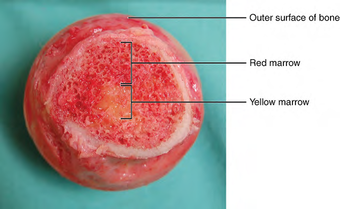
Figure 2. Head of Femur Showing Red and Yellow Marrow. The head of the femur contains both yellow in the center and red marrow surrounding it in the spongy bone with an outer surface of bone covering it. Yellow marrow stores fat. Red marrow is responsible for hematopoiesis.
CC BY: OpenStax College
The 206 bones that compose the adult skeleton are divided into five categories based on their shapes (Figure 3). Their shapes and their functions are related such that each categorical shape of bone has a distinct function.
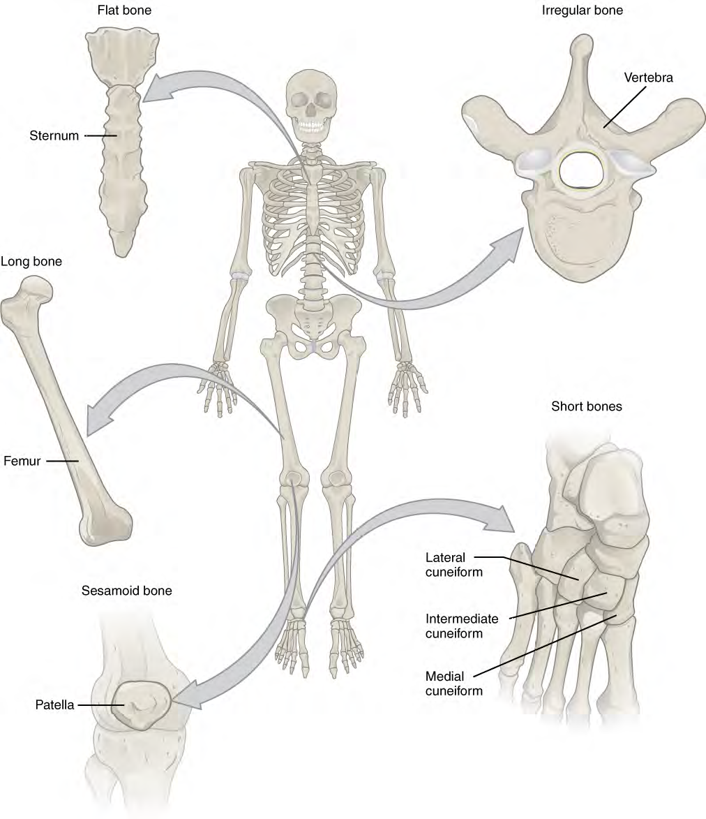
Figure 3. Bone Shape. This figure displays the skeleton as a whole with various bone shape examples enlarged to emphasize their form. These examples include the flat bone of the sternum, the irregular bones of the vertebrae, the long bone of the femur, the short bones only found in the feet and wrists, and the sesamoid (also called a round bone) patellar bone of the knee cap.
Long Bone
A long bone is longer than it is wide and is found in the appendages, such as the arm and hand. The long bones are not always obvious the hands and feet, however. Remember that they are longer than they are wide, and can be found in the metacarpals of the palm and phalanges which are the finger bones.
Short Bone
A short bone is one that is cube-like in shape, being approximately equal in length, width, and thickness. The ONLY short bones in the human skeleton are in the carpals of the wrists and the tarsals of the ankles. Short bones provide stability and support as well as some limited motion.
Flat Bone
The term "flat bone" is somewhat of a misnomer because, although a flat bone is typically thin, it is also often curved. Examples include the cranial (skull) bones, the scapulae (shoulder blades), the sternum (breastbone), and the ribs. Flat bones serve as points of attachment for muscles and often protect internal organs.
Irregular Bone
An irregular bone is one that does not have any easily characterized shape and therefore does not fit any other classification. These bones tend to have more complex shapes, like the vertebrae that support the spinal cord and protect it from compressive forces. Many facial bones, particularly the ones containing sinuses, are classified as irregular bones.
Sesamoid Bone
A sesamoid bone is a small, round bone that, as the name suggests, is shaped like a sesame seed. These bones form in tendons (the sheaths of tissue that connect bones to muscles) where a great deal of pressure is generated in a joint. The sesamoid bones protect tendons by helping them overcome compressive forces. The patellar bones of the knees are the only round bones found in common with every person.
| Bone Classification | Features | Functions | Examples |
|---|---|---|---|
|
Long |
Longer than wide |
Leverage |
Femur, metatarsals, ulna, phalanges |
|
Short |
Cube-like shape |
Provide stability while allowing for some movement |
Carpals, tarsals |
|
Flat |
Thin, curved |
Protects internal organs |
Sternum, ribs, cranial bones |
|
Irregular |
Unusual shape |
Protect organs |
Vertebrae, facial bones |
|
Sesamoid |
Small and round; inside tendons |
Protect tendons from compressed forces |
Patellar bones |
Video 1. View the video Bones- Shapes and Structures on YouTube (opens in new window)
Bone tissue (osseous tissue) differs greatly from other tissues in the body. Bone is hard and many of its functions depend on that characteristic hardness. Later discussions in this chapter will show that bone is also dynamic in that its shape adjusts to accommodate stresses. This section will examine the gross anatomy of bone first and then move on to its histology.
The structure of a long bone allows for the best visualization of all of the parts of a bone (Figure 4). A long bone has two parts: the diaphysis and the epiphysis. The diaphysis is the tubular shaft that runs between the proximal and distal ends of the bone. The hollow region in the diaphysis is called the medullary cavity, which is filled with yellow marrow. The walls of the diaphysis are composed of dense and hard compact bone.
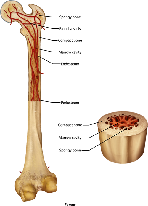
Figure 4. Anatomy of Long Bone. The femur bone here is cut longitudinally in half to reveal the inner marrow cavity of the shaft or diaphysis. The diaphysis contains the compact bone with a thin layer of spongy bone within that is lined with a membrane called the endosteum. At either end there is a head which is filled with spongy bone. Blood vessels run throughout the living bone, and it is covered by a periosteum.
The wider section at each end of the bone is called the epiphysis (plural = epiphyses), which is filled with spongy bone. Red marrow fills the spaces in the spongy bone. Each epiphysis meets the diaphysis at the metaphysis, the narrow area that contains the epiphyseal plate (growth plate), which is a layer of hyaline (transparent) cartilage in a growing bone. When the bone stops growing in early adulthood (approximately 18-21 years), the cartilage is replaced by osseous tissue and the epiphyseal plate becomes an epiphyseal line.
The medullary cavity has a delicate membranous lining called the endosteum (end- = "inside"; oste- = "bone"), where bone growth, repair, and remodeling occur. The outer surface of the bone is covered with a fibrous membrane called the periosteum (peri- = "around" or "surrounding"). The periosteum contains blood vessels, nerves, and lymphatic vessels that nourish compact bone. Tendons and ligaments also attach to bones at the periosteum.The periosteum covers the entire outer surface except where the epiphyses meet other bones to form joints (Figure 5). In this region, the epiphyses are covered with articular cartilage, a thin layer of hyaline cartilage that reduces friction and acts as a shock absorber.

Figure 5. Compact and Spongy Bone. This small section of bone is magnified to show the way compact bone forms osteons in cylinders throughout the length of the bone (this is from the outer part of the shaft as seen in Figure 4). The groups of concentric circles of compact bone, called lamellae, are the osteons that are considered the functional unit of compact bone. Within a central canal of the osteon are capillaries and small veins. These blood vessels came into the bone through perforating canals lying horizontal in the compact bone. In addition to the concentric lamellae, there are also sections of interstitial lamellae seen between the osteons as well as circumferential lamellae that lie beneath the outer periosteal layer.
Flat bones, like those of the cranium, consist of a layer of diploe (spongy bone), lined on either side by a layer of compact bone (Figure 6). The two layers of compact bone and the interior spongy bone work together to protect the internal organs. If the outer layer of a cranial bone fractures, the brain is still protected by the intact inner layer.
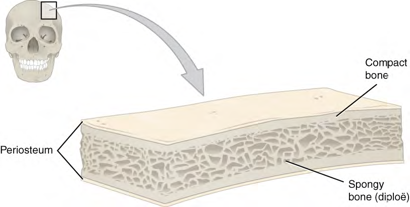
Figure 6. A cross-section of a flat bone shows the spongy bone on the inside with the compact bone lining it on either side, like a sandwich of spongy, diploe, bone between two layers of compact bone.
CC BY: OpenStax College
The surfaces of bones have various bone markings for attachments of tendons or for articulations to other bones (Figure 7). These will vary depending on location and function of the bone marking. Bone markings fall into two general categories: projections and depressions. A projection is an area of a bone that projects above the surface of the bone, which often articulates with a bone depression on a facing bone. Other projections will hold tendons and ligaments. Depressions and openings allow blood vessels and nerves to enter the bone, or allow an articulation with another bone. On your own, fill in a table like the one below of the primary markings.
| Marking | Description | Example |
|---|---|---|
|
Head |
Prominent, rounded surface at the end of a bone |
Blank |
|
Condyle |
Smooth rounded surface for an articulation |
Blank |
|
Spine |
Sharp process |
Blank |
|
Tubercle |
Small, rounded process |
Blank |
|
Tuberosity |
Rough surface where a tendon attaches |
Blank |
|
Trochanter |
Large rounded process for muscle attachment on the femur |
Blank |
|
Crest |
Ridge |
Blank |
|
Process |
Prominent feature emerging from a bone |
Blank |
|
Foramen |
Hole through a bone |
Blank |
|
Fossa |
Elongated basin |
Blank |
|
Fovea |
Small pit |
Blank |
|
Fissure |
Slit through a bone |
Blank |
|
Meatus |
Opening into a canal |
Blank |
|
Sinus |
Air-filled space in a bone |
Blank |
Download the Table 2 Bone Markings Sudent Table PDF file.
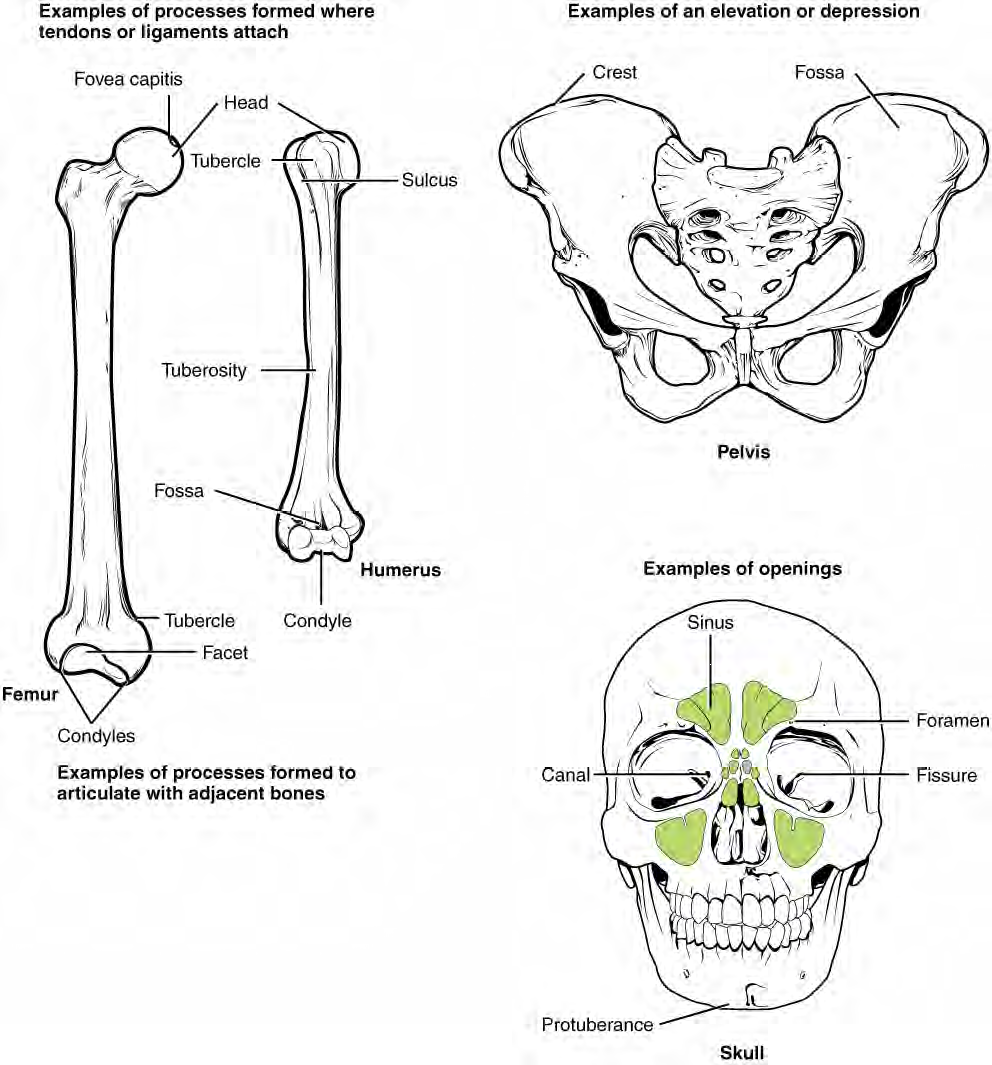
Figure 7. Bone Markings. The general bone markings include processes that extend from the bone in various ways and depressions which are indented into the bone or form holes (foramen). The examples of processes here include the head, tubercle, tuberosity, condyle, protuberance and crest. The examples of depressions include fossa, canal, fissure, and a hole or foramen.
CC BY: OpenStax College
Like other connective tissues, bone, or osseous tissue, contains specialized cells surrounded by an extracellular matrix that contains fibers and various mineral salts. These salt crystals form when calcium phosphate [Ca3(PO4)2] and calcium carbonate [Ca(OH)2] combine to create hydroxyapatite [Ca10(PO4)6(OH)2] (hī-drok-sē-AP-a-tīt), which incorporates other inorganic salts like magnesium hydroxide, fluoride, and sulfate as it crystallizes, or calcifies, on the collagen fibers. The hydroxyapatite crystals give bones their hardness and strength, while the collagen fibers give them flexibility so that they are not brittle. Although bone cells compose a small amount of the bone volume, they are crucial to the function of bones. Four types of cells are found within bone tissue:
The osteoblast is the bone cell responsible for building new bone and is found in the growing portions of bone, including the periosteum and endosteum. Osteoblasts, which do not divide through mitosis , synthesize collagen and secrete the matrix and calcium salts. As the secreted matrix surrounding the osteoblast calcifies, the osteoblast become trapped within it; as a result, it changes in structure and becomes an osteocyte, the primary cell of mature bone and the most common type of bone cell. Each osteocyte is located in a space called a lacuna and is surrounded by bone tissue. Osteocytes maintain the mineral concentration of the matrix via the secretion of enzymes. Like osteoblasts, osteocytes lack mitotic activity. They can communicate with each other and receive nutrients via long cytoplasmic processes that extend through canaliculi (singular = canaliculus), little, canal-like channels within the bone matrix.
If osteoblasts and osteocytes are incapable of mitosis, then how are they replenished when old ones die? The answer lies in the properties of a third category of bone cell, the osteogenic cell. These osteogenic cells are stem cells with high mitotic activity and they are the only bone cells that divide. Immature osteogenic cells are found in the deep layers of the periosteum and the marrow. They differentiate and develop into osteoblasts.
The dynamic nature of bone means that new tissue is constantly formed, and old, injured, or unnecessary bone is dissolved for repair or for calcium release. The cell responsible for bone resorption (meaning the breakdown or cutting away of bone) is the osteoclast. They are found on bone surfaces, are multinucleated, and originate from monocytes and macrophages, two types of white blood cells, not from osteogenic cells. Osteoclasts are continually breaking down old bone while osteoblasts are continually forming new bone. The continuous balance between osteoblasts building up bone and osteoclasts cutting it away is responsible for the constant but subtle reshaping of bone, so that over about 10 years you have a new skeleton.
Use the word "builder" to remember the function of Osteoblasts and "cutter" to remember that Osteoclasts cut away at the bone. Here is a table to fill out from the information above:
| Cell Type | Function | Location |
|---|---|---|
|
Osteogenic |
Blank |
Blank |
|
Osteoblast |
Blank |
Blank |
|
Osteocyte |
Blank |
Blank |
|
Osteoclast |
Blank |
Blank |

The differences between compact and spongy bone are best explored via their histology. Most bones contain compact and spongy osseous tissue, but their distribution and concentration vary based on the bone's overall function. Compact bone is dense so that it can withstand compressive forces, while spongy (cancellous) bone has open spaces and supports shifts in weight distribution.
Figure 8. Compact and Spongy Bone. Shows cyllindrical concentric lamellae going vertical to withstand compressive forces
Compact bone is the denser, stronger of the two types of bone tissue (Figure 8). It can be found under the periosteum and in the diaphyses of long bones, where it provides support and protection. The microscopic structural unit of compact bone is called an osteon, or Haversian system. Each osteon is composed of concentric rings of calcified matrix called lamellae (singular = lamella). Running down the center of each osteon is the central canal, which contains blood vessels, nerves, and lymphatic vessels. These vessels and nerves branch off at right angles through a perforating canal, also known as Volkmann's canals, to extend to the periosteum and endosteum. The osteocytes are located inside spaces called lacunae (singular = lacuna), found at the borders of adjacent lamellae. As described earlier, canaliculi connect with the canaliculi of other lacunae and eventually with the central canal (Figure 9). This system allows nutrients to be transported to the osteocytes and wastes to be removed from them, as they maintain the matrix.
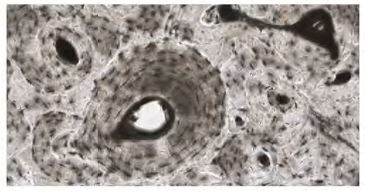
Figure 9. In this micrograph of the osteon, you can clearly see the concentric lamellae and central canals in addition to the osteocytes in lacunae. LM × 40
(Micrograph provided by the Regents of University of Michigan Medical School c 2012) from Open Stax.
Like compact bone, spongy bone, also known as cancellous bone, contains osteocytes housed in lacunae, but they are not arranged in concentric circles. Instead, the lacunae and osteocytes are found in a lattice-like network of matrix spikes called trabeculae (singular = trabecula) (Figure 8). The trabeculae may appear to be a random network, but each trabecula forms along lines of stress to provide strength to the bone. The spaces of the trabeculated network provide balance to the dense and heavy compact bone by making bones lighter so that muscles can move them more easily. In addition, the spaces in some spongy bones contain red marrow, protected by the trabeculae, where hematopoiesis occurs.


Except where otherwise noted, this work by The Community College Consortium for Bioscience Credentials is licensed under a Creative Commons Attribution 4.0 International License.
Text from BioBook licensed under CC BY NC SA and Boundless Biology Open Textbook licensed under CC BY SA..
Other text from OpenStaxCollege licensed under CC BY 3.0. Modified by Alice Rudolph, M.A. and Amy Bauguess, M.S. for c3bc.
Instructional Design by Courtney A. Harrington, Ph.D., Helen Dollyhite, M.A. and Caroline Smith, M.A. for c3bc.
Media by Brittany Clark and Antonio Davis for c3bc.
This product was funded by a grant awarded by the U.S. Department of Labor's Employment and Training Administration. The product was created by the grantee and does not necessarily reflect the official position of the U.S. Department of Labor. The Department of Labor makes no guarantees, warranties, or assurances of any kind, express or implied, with respect to such information, including any information on linked sites and including, but not limited to, accuracy of the information or its completeness, timeliness, usefulness, adequacy, continued availability, or ownership.