Factors that Affect Skin Functions
Integumentary System
Impacting the Function of Skin
Objectives
- Students will be able to describe the changes that occur in the integumentary system during the aging process.
- Students will be able to discuss several common diseases, disorders, and injuries that affect the integumentary system.
- Students will be able to explain treatments for some common diseases, disorders, and injuries of the integumentary system.
Skin Pigment Disorders
Albinism is a congenital disorder due to the defect of an enzyme that helps make melanin; these individuals tend to need more protection from UV radiation, since they are more prone to sunburns and skin cancer; they also tend to be more sensitive to light and have vision problems due to the lack of pigmentation on the retinal wall; treatment of this disorder involves addressing the symptoms, such as limiting UV light exposure to the skin and eyes, etc. Find some more information about this disorder and its worldwide cultural impacts here:
Video 1. Albinism (opens Youtube in new window)
CC BY: OpenStax College via ABC News
Vitiligo is a chronic disorder that causes depigmentation patches in the skin; the melanocytes in certain areas lose their ability to produce melanin; see normal melanocyte layering in Figure 1.
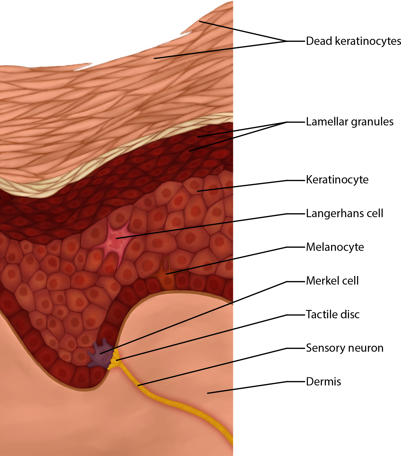
Figure 1. Epidermal Layers; note the melanocytes and think about their contribution; melanocytes can be found in the stratum basale, but they have cellular extensions that go into the stratum spinosum above, spreading their pigments and color into the surrounding areas of the skin.
Other changes in the appearance of skin coloration can be indicative of diseases associated with other body systems. Liver disease or liver cancer can cause the accumulation of bile and the yellow pigment bilirubin, leading to the skin appearing yellow or jaundiced (jaune is the French word for "yellow"). Addison's disease can stimulate the release of excess amounts of adrenocorticotropic hormone (ACTH), which can give the skin a deep bronze color. A sudden drop in oxygenation can affect skin color, causing the skin to initially turn ashen (white). With a prolonged reduction in oxygen levels, dark red deoxyhemoglobin becomes dominant in the blood, making the skin appear blue, a condition referred to as cyanosis (kyanos is the Greek word for "blue"). This happens when the oxygen supply is restricted, as when someone is experiencing difficulty in breathing because of asthma or a heart attack. However, in these cases the effect on skin color has nothing do with the skin's pigmentation.
Vitamin D Comes From the Skin (not the sun)
The epidermal layer of human skin synthesizes vitamin D when exposed to UV radiation. In the presence of sunlight, a form of vitamin D3 called cholecalciferol is synthesized from cholesterol in the skin. Vitamin D is essential for normal absorption of calcium and phosphorous, which are required for healthy bones among other needs. The absence of sun exposure can lead to a lack of vitamin D in the body, leading to a condition called rickets, a painful condition in children where the bones are misshapen due to a lack of calcium, causing bow-leggedness. Elderly individuals who suffer from vitamin D deficiency can develop a condition called osteomalacia, a softening of the bones. In present day society, vitamin D is added as a supplement to many foods, including milk and orange juice, compensating for the need for sun exposure.
In addition to its essential role in bone health, vitamin D is essential for general immunity against bacterial, viral, and fungal infections. Recent studies are also finding a link between insufficient vitamin D and cancer.
Overexposure to UV radiation damages DNA, which can lead to the formation of cancerous skin lesions. Although melanin offers some protection against DNA damage from the sun, often it is not enough. Basal cell carcinoma is a form of cancer that affects the mitotically active stem cells in the stratum basale of the epidermis. It is the most common of all cancers that occur in the United States and is frequently found on the head, neck, arms, and back, which are areas that are most susceptible to long-term sun exposure. The appearance of this cancer will vary depending on the individual and the form of cancer. The lesion can turn hands from a shiny pearl like area to a red eczema like area. Squamous cell carcinoma is a cancer that affects the keratinocytes of the stratum spinosum and presents as lesions commonly found on the scalp, ears, and hands. These lesions also vary in appearance but tend to be scaly, crusty, wart like, and possibly bleeding. It is the second most common skin cancer. A melanoma is a cancer characterized by the uncontrolled growth of melanocytes, the pigment-producing cells in the epidermis. Typically, a melanoma develops from a mole. It is the most fatal of all skin cancers, as it is highly metastatic and can be difficult to detect before it has spread to other organs. To identify a melanoma lesion the ABCDEs are used - any of these conditions present in a mole can indicate melanoma.
A = Asymmetry, if you draw a line through the center of the mole and the 2 sides do not match that means the mole is asymmetrical and is unlike a benign mole.
B = Border, instead of a typical rounded border you see scalloped edges and it is uneven the mole may be cancerous.
C = Color, if the mole is not uniform or homogeneous in color it may be cancerous.
D = Diameter, if the diameter of the mole is greater than 6mm it may be cancerous.
E = Evolving, or changing over time, may indicate a cancerous mole.
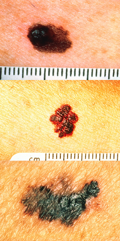
Figure 2. Melanoma; Note the changes in all ABCDEs described above. You can see they are asymmetrical, they uneven color and the uneven borders of the moles. You can see the width is larger than 6mm.
Image modified from a larger version by the Skin Cancer Foundation.
CCBY: OpenStax
Aging and the Skin
As we age, everything about the skin seems to decrease. The regeneration ability of the skin reduces, causing thinner skin. The accessory structures also have lowered activity, generating thinner hair and nails, and reduced amounts of sebum and sweat. A reduced sweating ability can cause some elderly to be intolerant to extreme heat, so be careful to carry water with you. Other cells in the skin, such as melanocytes and immunological cells also become less active, leading to a paler skin tone, white hair and lowered immunity. Wrinkling of the skin occurs due to breakdown of its structure, which results from decreased collagen and elastin production in the dermis, weakening of muscles lying under the skin, and the inability of the skin to retain adequate moisture.
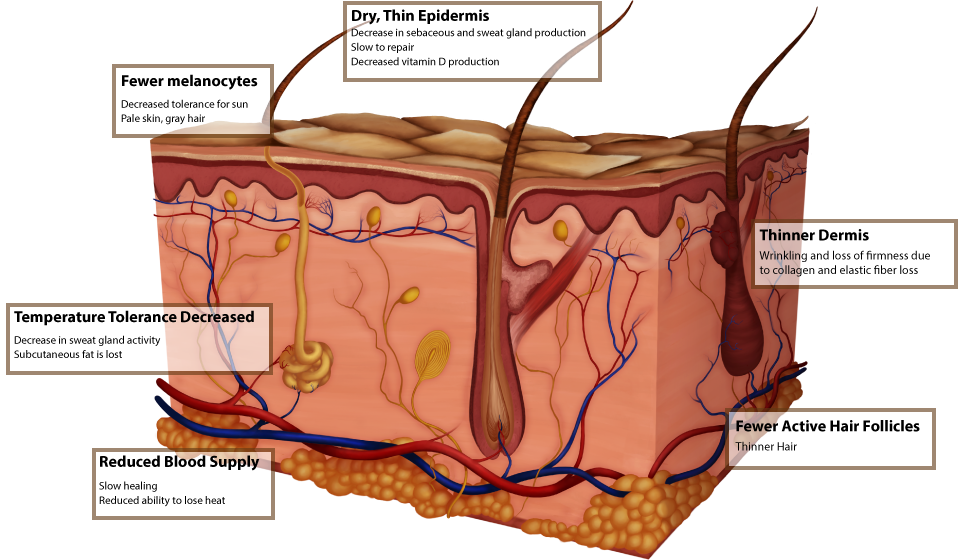
Figure 3. Aging Skin. In aging skin the epidermis becomes dry and thin, wrinkling occurs due to collagen and elastic fiber loss, temperature tolerance is decreased, there are fewer melanocytes, reduced blood supply and fewer active hair follicles.
Skin Repair
Two kinds of wound-healing processes can occur, depending on how deep the injury is in the skin. Epidermal wound healing occurs following superficial wounds. The epidermal layer will typically return to normal function. An example of an epidermal wound is a paper cut. Deep wound healing occurs when an injury extends to the dermis and subcutaneous layer. Loss of some function and development of scar tissue is the rule. An example of a deep wound would be injuries that leave scars whether they are from surgery, imbalance, or a lack of common sense. The skin does have a lot of collagen in it (lab models of dermis often have lots of noticable pink fibers), which needs Vitamin C to develop its functioning structure. Smoking interferes with collagen's proper development and can slow down would healing.
Burns
A burn is tissue damage caused by excessive heat, electricity, radioactivity, or chemicals that denature (break down) the proteins in the skin cells. Burns destroy some of the skin's important contributions to homeostasis — protection against microbial invasion, desiccation, thermoregulation, etc. Burns are graded according to their severity.
A first-degree burn damages only through the epidermis (first main layer) like a sunburn.
A second-degree burn destroys the epidermis and part of the dermis causing redness, blister formation, edema, pain, and some skin functions are lost.
A third-degree burn is a full-thickness burn (destroys the epidermis, dermis, and subcutaneous layer). Most skin functions are lost, and the region is numb because sensory nerve endings have been destroyed.
Burns are sometimes measured in terms of the size of the total surface area affected. This is referred to as the "rule of nines," which associates specific anatomical areas with a percentage that is a factor of nine. Each arm is 9% of the body (front and back), the front of each leg is 9%, so each leg is 18%. The torso is a total of 36%. The size of the burn will constitute what the nurse and doctor should do to treat or help the patient, i.e.: the burn victim will need wound care and fluid resuscitation (since risks may include hypovolemia). Destruction of skin layers is usually referred to as a partial-/full-thickness burn, which will require grafting from another area of the patient's body. Depending on the type of burn, often a pressure cuff is worn for a few months after the wound has closed.
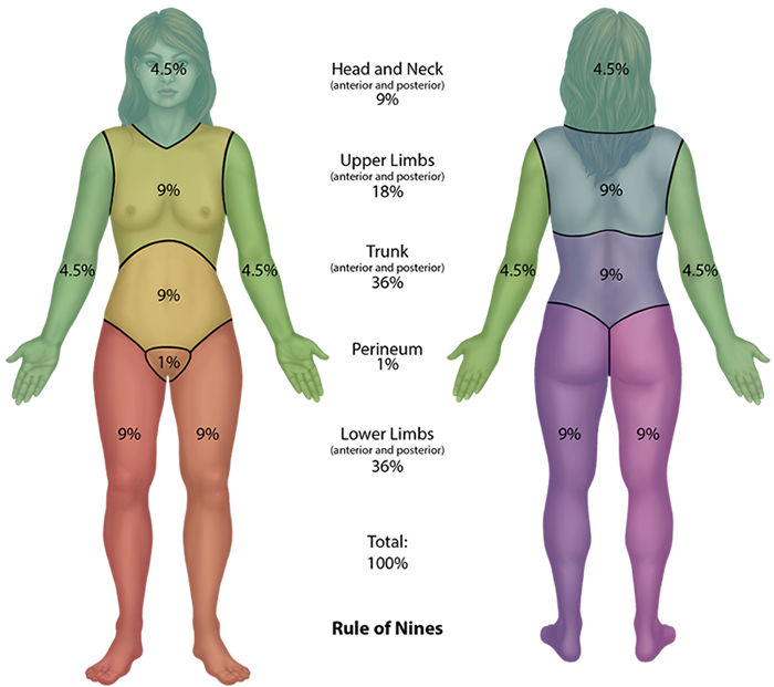
Figure 4. Rule of Nines. The body is divided into areas to determine the extent of a burn. The legs are 9% each anteriorly and 9% posteriorly (36% total), the arms are 4.5% each, the back is 18% and the anterior trunk is 18%, the head and neck are 9%, and the perineum covers 1%.
Scars and Keloids
Most cuts or wounds, with the exception of ones that only scratch the surface (the epidermis), lead to scar formation. A scar is collagen-rich skin formed after the process of wound healing that differs from normal skin. Scarring occurs in cases in which there is repair of skin damage, but the skin fails to regenerate the original skin structure. Fibroblasts generate scar tissue in the form of collagen, and the bulk of repair is due to the basket-weave pattern generated by collagen fibers and does not result in regeneration of the typical cellular structure of skin. Instead, the tissue is fibrous in nature and does not allow for the regeneration of accessory structures, such as hair follicles, sweat glands, or sebaceous glands. Scarring of skin after wound healing is a natural process and does not need to be treated further. Sometimes, there is an overproduction of scar tissue, because the process of collagen formation does not stop when the wound is healed; this results in the formation of a raised or hypertrophic scar called a keloid.
Bedsores and Stretch Marks
Skin and its underlying tissue can be affected by excessive pressure. One example of this is called a bedsore. Bedsores, also called decubitis ulcers, are caused by constant, long-term, unrelieved pressure on certain body parts that are bony, reducing blood flow to the area and leading to necrosis (tissue death). Bedsores are most common in elderly patients who have debilitating conditions that cause them to be immobile. Most hospitals and long-term care facilities have the practice of turning the patients every few hours to prevent the incidence of bedsores. If left untreated, bedsores can be fatal if they become infected.
The skin can also be affected by pressure associated with rapid growth. A stretch mark results when the dermis is stretched beyond its limits of elasticity, as the skin stretches to accommodate the excess pressure. Stretch marks usually accompany rapid weight gain during puberty and pregnancy. They initially have a reddish hue, but lighten over time.
Self-Assessment

Glossary
- Addison's Disease
- A rare hormonal disease that can give the skin a deep bronze color due to excess adrenocorticotropic hormone (ACTH).
- Albinism
- genetic disorder that affects the skin, in which there is no melanin production.
- Basal Cell Carcinoma
- cancer that originates from basal cells in the epidermis of the skin.
- Bedsore
- tissue damage caused by unrelieved pressure on body parts that are bony.
- Cyanosis
- with a prolonged reduction in oxygen levels, dark red deoxyhemoglobin becomes dominant in the blood, making the skin appear blue.
- First-Degree Burn
- damages only through the epidermis (first main layer) like a sunburn.
- Jaundiced
- An accumulation of bile and the yellow pigment bilirubin that leads the skin to appear yellow; often caused by liver disease or liver cancer.
- Keloid
- a raised or hypertrophic scar.
- Melanoma
- type of skin cancer that originates from the melanocytes of the skin.
- Rickets
- disease in children caused by vitamin D deficiency, which leads to the weakening of bones.
- Scar
- collagen-rich skin formed after the process of wound healing that differs from normal skin.
- Second-Degree Burn
- destroys the epidermis and part of the dermis causing redness, blister formation, edema, pain, and some skin functions are lost.
- Squamous Cell Carcinoma
- type of skin cancer that originates from the stratum spinosum of the epidermis.
- Stretch Mark
- a mark left when skin is stretched beyond the limits of its elasticity.
- Third-Degree Burn
- is a full-thickness burn (destroys the epidermis, dermis, and subcutaneous layer). Most skin functions are lost, and the region is numb because sensory nerve endings have been destroyed.
- Vitiligo
- skin condition in which melanocytes in certain areas lose the ability to produce melanin, possibly due an autoimmune reaction that leads to loss of color in patches.
Grant and Other Information

Except where otherwise noted, this work by The Community College Consortium for Bioscience Credentials is licensed under a Creative Commons Attribution 4.0 International License.
Text from BioBook licensed under CC BY NC SA and Boundless Biology Open Textbook licensed under CC BY SA.
Other text from OpenStaxCollege licensed under CC BY 3.0. Modified by Alice Rudolph, M.A. for c3bc.
Instructional Design by Courtney A. Harrington, Ph.D., Helen Dollyhite, M.A. and Caroline Smith, M.A. for c3bc.
Media by Brittany Clark and Antonio Davis for c3bc.
This product was funded by a grant awarded by the U.S. Department of Labor's Employment and Training Administration. The product was created by the grantee and does not necessarily reflect the official position of the U.S. Department of Labor. The Department of Labor makes no guarantees, warranties, or assurances of any kind, express or implied, with respect to such information, including any information on linked sites and including, but not limited to, accuracy of the information or its completeness, timeliness, usefulness, adequacy, continued availability, or ownership.