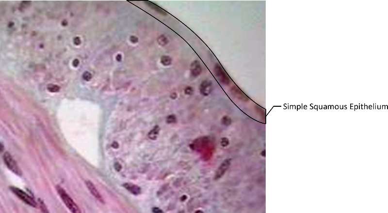
Figure 1. Cross section of small intestine (dyed purple for ease of visabilty). Simple Squamous Epithelium is outlined in black in the upper right corner.
Characteristics of Primary Tissue Types
Anatomy & Physiology I
You have learned about the different levels of organization in living things. You may recall that In multicelular organisms, groups of similar cells that work together to perform a particular function are called tissues. Animals are multicellular organisms. In animals, groups of similar cells work together to perform a particular function. Most animals have tissues that are very specialized.
There are four basic categories of tissues; epithelial tissue, muscle tissue, connective tissue, and nervous tissue. There are many specific types of tissues within each category. For example, we will observe five types of epithelium. Tissues in each category do share some general functions.
The following video introduces tissues.
Video 1. View the Tissues video on YouTube (opens in a new window).
Epithelial tissues line and cover body surfaces. For example, they line the stomach, line the ducts of glands, and cover our body. Because of the location of epithelial tissues, each type has a free surface (that faces open space) and a basement membrane (where they are connected to the body). Epithelial tissues are used for protection, absorption, secretion, filtration, and diffusion. The cells in epithelial tissue are very close together. The cells may be found in layers (stratified). Alternatively, the tissue may be only one cell layer thick (simple). Epithelium does not contain blood vessels, the tissue must get its nutrients from underlying connective tissue. We will consider six types of epithelium. Each type has a different structure and function. Students should be able to find and identify these tissues and their characteristic structure.
| Tissue | Structure | Function | Representative Locations |
|---|---|---|---|
|
Simple Squamous Epithelium |
very thin, flat cells, one cell layer thick |
diffusion, osmosis, filtration |
air sacs in lungs, line blood vessels, capillaries, intestine surface |
|
Simple Columnar Epithelium |
single layer of very tall cells, some are goblet cells |
protection, secretion, absorption |
lines stomach and intestines, uterus |
|
Pseudostratified Columnar Epithelium with cilia |
simple tissue but varying location of nuclei gives the appearance of striations, goblet cells, cilia |
protection, secretion, movement of cells and particles |
trachea, fallopian tubes |
|
Simple Cuboidal Epithelium |
simple tissue, cells are cube shaped with nucleus in the middle |
secretion, absorption |
lines ducts of glands, lines kidney tubules |
|
Stratified Squamous Epithelium |
many layers of cells with the cells towards the surface being flat and the deeper cells more cuboidal |
protection |
skin - keratinized, lining of oral cavity, vagina - nonkeratinized |
|
Transitional Epithelium |
appears to have many layers when the tissue is not stretched; appears to have fewer layers when the tissue is stretched |
as the bladder empties and fills, it's able to adapt. |
lines urinary bladder, urethra |
The following video also discusses the types of epithelial tissues.
Video 2. View the Epithelial Tissues video on YouTube (opens in a new window)
Simple squamous epithelium is a very thin, flat-celled tissue. Because the cells are thin, this tissue is found where transport of chemical compounds needs to be fast. For example, this tissue is found in alveoli in your lungs, allowing oxygen to diffuse quickly.
The following video discusses simple squamous epithelium.
Video 3. View the Simple Squamous Epithelium video on youTube (opens in a new window).
Figure 1. Cross section of small intestine (dyed purple for ease of visabilty). Simple Squamous Epithelium is outlined in black in the upper right corner.
The word simple tells you that simple columnar epithelium is composed of a single layer of cells. They are very tall and column shaped. Some of the cells are goblet cells, which aid in mucous secretion. Simple columnar epithelium is found lining the stomach, intestines and uterus. It functions to protect, secrete and absorb.
The following video discusses simple columnar epithelium.
Video 4. View the Simple Columnar Epithelium video on YouTube (opens in a new window)
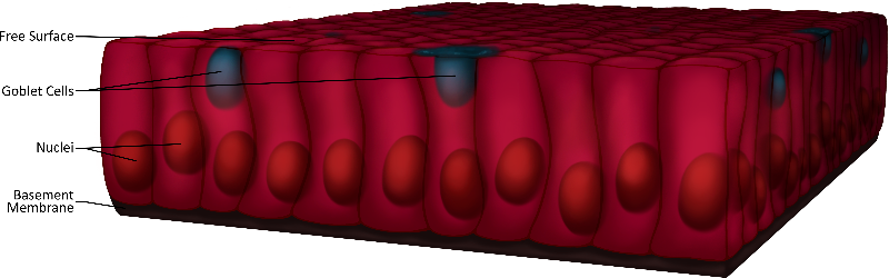
Figure 2. Cross section of simple columnar epithelium; The free surface, goblet cells, nuclei and basement membrane are labeled; note the column-shaped epithelial cells.
The prefix psuedo indicates that the tissue is not truly stratified. The varying locations of nuclei in this tissue gives the appearance of stratification, but the tissue is a single layer, simple tissue. Pseudostratified columnar epithelium with cilia also contains goblet cells, and as the name implies, has cilia. These aid in secretion and movement of cells and particles. This tissue is found in places that small particles or cells may need to be moved along, like the lining of the trachea and the fallopian tubes. The goblet cells secrete mucous which lubricates and protects the free surface.
The following video discusses psuedostratified columnar epithelium with cilia.
Video 5. View the Psuedostratified Columnar Epithelium video on YouTube (opens in a new window)
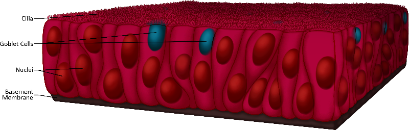
Figure 3. Cross section of pseudostratified columnar epithelium; The ciliated surface, goblet cells, nuclei and basement membrane are labeled; note the "semi-layering" of the column-shaped epithelial cells.
This tissue is made up of a single layer of cube shaped cells with their nuclei located in the middle. The functions include secretion and absorption and are found lining the ducts of glands, and kidney tubules.
The following video discusses simple cuboidal epithelium.
Video 6. View Simple Cuboidal Epithelium video on YouTube (opens in a new window).
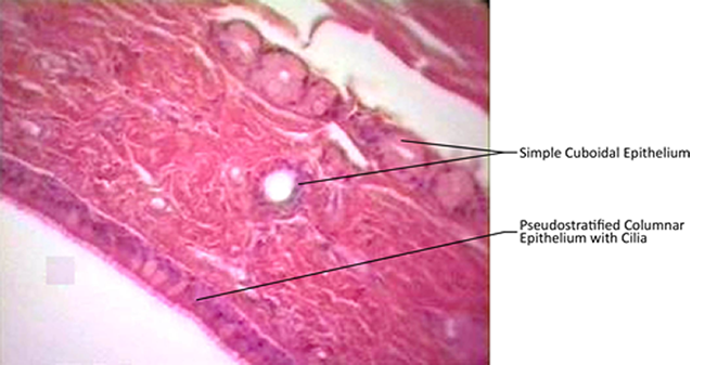
Figure 4. Micrographic trachea cross-section (dyed purple for ease of visabilty) with different types of epithelium labeled.
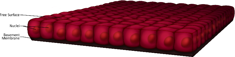
Figure 5. Simple cuboidal epithlium with free surface, nuclei and basement membrane labeled; note the cube-shaped cells.
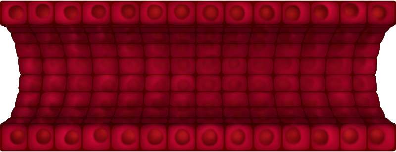
Figure 6. Longitudinal cross-section of a duct made of simple cuboidal epithelial tissue.
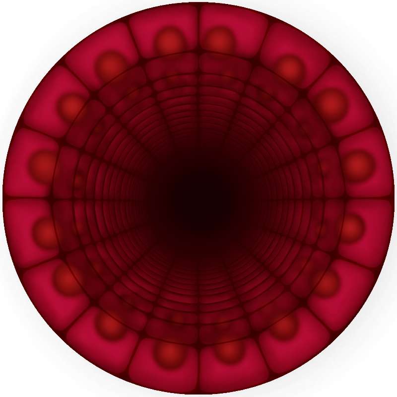
Figure 7. Cross-section of a duct made of simple cuboidal epithelial tissue.
Stratified squamous epithelium has many layers of cells. The cells toward the free surface are flatter and those closer to the basement membrane are more cube-shaped. The main function of this tissue is protection. There are two kinds of stratified squamous epithelium: keratinized and nonkeratinized. The keratinized tissue is very tough because the cells contain the protein keratin. The cells in the outermost layer are dead, heavily keratinized cells. Keratininzed stratified squamous epithelium is found in your skin. Non keratinized stratified squamous epithelium is located in places such as the lining of the oral cavity and the rectum. This tissue is softer but its many layers of cells still serves to protect the underlying tissues. We will learn more about this tissue when we study the skin.
The following video discusses stratified squamous epithelial tissue.
Video 7. View the Stratified Squamous Epithelial Tissue video on YouTube (opens in a new window).
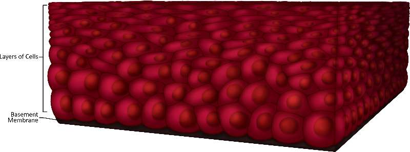
Figure 8. Cross-section of stratified squamous epitheial tissue; note the stacking of cells and the impact that has on their shape.
Transitional epithelium appears to have few layers when it is stretched and many layers when it is not. It's found lining the bladder and when the bladder is full, that's when the tissue is stretched and appears to only have a few layers. When the bladder is emptied, the tissue appears to have many layers. Compare the two images below to see the difference.
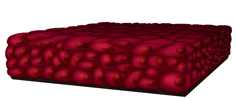
Figure 9. The image represents the changes that transitional epithelium tissues go through. Note the compactness of the cells.
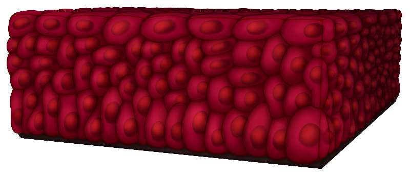
Figure 10. Represent the changes that transitional epithelium tissues go through. Note the the inflated-seeming cells.
The following video discusses transitional epithelium.
Video 8. View theTransitional Epithelium video on YouTube (opens in a new window).
Epithelial glands are an example of simple organs. Glands that secrete their contents directly into the blood are called endocrine glands. Glands that secrete their contents into a lumen or duct are called exocrine glands. We will look at some common types of exocrine glands (endocrine glands are studied later).
Exocrine Glands secrete products through ducts to the surface of the skin or into the lumen of a hollow organ (examples include the sweat ducts or pancreatic ducts). Secretions of the exocrine gland include mucus, sweat, oil, earwax, saliva, and digestive enzymes. Examples of exocrine glands are sudoriferous (sweat) glands.
These multicellular glands are categorized according to function based on the way the gland secretes its product from cell's cytoplasm to the outside environment. The three forms of secretion are: Merocrine, Apocrine and Holocrine.
Exocrine Gland Secretion
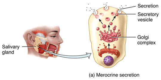
Figure 11: Merocrine secretion. Our salivary glands secrete digestive enzymes by the merocrine method. You can see a cell in this figure that shows the Golgi complex forming secretory vesicles of salivary enzymes, which merge with the outside cell membrane to release them into the mouth for digesting carbohydrates. No part of the cell is lost during merocrine secretion, since secretion happens by this exocytosis method.
Merocrine secretion is the most common manner of secretion (you can remember merocrine and most both with the first letter "m"). The gland releases its product by exocytosis and no part of the gland is lost or damaged. Your salivary glands secrete this way (Figure 10). Also, our sweat glands secrete by the merocrine method, too. Any type of sweat that develops a strong odor only does so because it comes into contact with bacteria on the surface of the skin, as at the axillary area.
Apocrine gland secretions leave via a group of vesicles that accumulate at the top or apex of the cell, thus its name. The end of the cell breaks off once it is filled with these vesicles leaving a milky, viscous odorless fluid. Apocrine glands are not those of the axillary regions (those are still the merocrine types of glandular secretion), but are found in the mammary glands.
Holocrine secretions are produced when the plasma membrane breaks, releasing the entire cellular contents into the lumen and killing the cell (cells are replaced by rapid division of stem cells.) The sebaceous gland is an example of a holocrine gland, because its secretion (sebum) is released with remnants of dead cells.
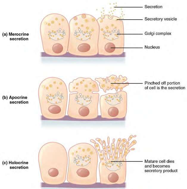
Figure 12: Modes of Glandular Secretion. (a) In merocrine secretion, the cell remains intact using vesicles and exocytosis to release secretions. (b) In apocrine secretion, the apical portion of the cell is released, as well as the material to be secreted. An example is milk secretion from the mammary glands (c) In holocrine secretion, the cell is destroyed as it releases its product and the cell itself becomes part of the secretion; the whole cell dies, so new ones form from stem cells, as happens in the skin's sebaceous glands.
Connective Tissue is found throughout the organism. It holds structures together, provides support and protection, gives structures a flexible and/or elastic quality, and absorbs shock. Some tissues are also involved with storage. The cells in connective tissue are far apart. The matrix is the material found between the cells. The matrix contains fibers and a ground substance.
There are three categories of connective tissue cells. A -blast is a cell that secretes the matrix. A -clast is a cell that destroys the matrix. A -cyte is a cell that maintains the matrix.
There are three types of fibers that can be found in connective tissues. Elastic fibers are thin fibers that have the ability to stretch and return to their original shape. They are made of the protein elastin. Collagenous fibers are thick fibers made of the protein collagen. They are very tough and have a high tensile strength. Reticular fibers are thin fibers made of collagen. They give support to solid organs such as the liver.
|
Tissue |
Structure |
Function |
Representative Location
|
|
Loose Connective Tissue or Areolar |
Both collagenous and elastic fibers suspended in a thick, fluid ground substance with scattered fibroblasts |
Binds organs together, |
Beneath the skin, between muscles |
|
Adipose tissue |
Large cells with a fat storage area, nucleus is pushed to the side by stored fat |
store fat, protection, insulation |
under skin, behind eyes, around kidneys and heart |
|
Fibrous or Dense Connective Tissue |
Thick, dense collagenous fibers with scattered fibroblasts |
strong support |
tendons, ligaments |
|
Hyaline Cartilage |
Chondrocytes in lacuna, matrix of chondrin |
flexible support, protection |
ends of long bones, end of nose, trachea |
|
Blood |
Red Blood Cells, White Blood Cells, and platelets in liquid matrix (plasma) |
transport, protection |
blood vessels |
|
Bone |
Osteocytes in lacuna, matrix of calcium salts, central canal |
support, protection |
bones |
The following video gives an overview of connective tissue.
Video 9. View the Connective Tissue video on YouTube (opens in a new window)
Loose connective tissue binds organs together, it is found between different layers of tissues and between organs. For example, under the skin, and between muscles. It is made up of collagenous and elastic fibers suspended in a thick, ground substance with fibroblasts scattered throughout.
The following video discusses loose connective tissue.
Video 10. View the Connective Tissues video on YouTube (opens in a new window)
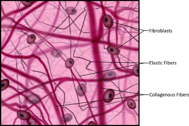
Figure 13. Illustration of connective tissue, often intersected by lines of various widths, representing elastic and collagenous fibers, which are produced in the simple oval-shaped fibroblasts also pictured.
Adipose tissue provides protection, insulation and fat storage in large cells where the nucleus is pushed to the side by large areas for fat storage. It's found under the skin, behind the eyes, surrounding the heart and kidneys.
The following video discuses adipose tissue.
Video 11. View the Adipose Tissue video on YouTube (opens in a new window)
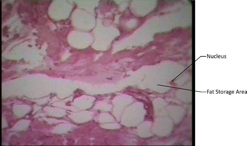
Figure 14. A micrograph dyed purple for ease of visabilty.
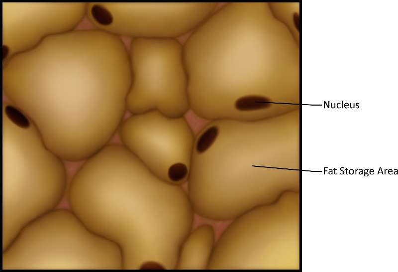
Figure 15. A close-up illustration; note the globular shape of the fat cells.
Hyaline cartilage is located in places such as at the ends of long bones, the end of the nose and in the trachea. It is made up of chondrocytes located in lacuna (which means chamber) in a matrix of chondrin. It's purpose is to provide protection and support.
The following video discusses hyaline cartilage.
Video 12. View the Hyaline Carilage video on YouTube (opens in a new window)
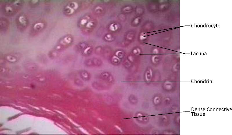
Figure 16. A cross-sections of hyaline cartilage in the trachea where the actual micrograph has been dyed purple for ease of visabilty.
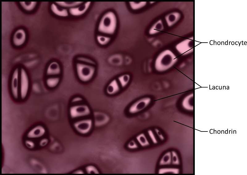
Figure 17. An illustration of hyaline cartilage in the trachea where the actual micrograph has been dyed purple for ease of visabilty.
Blood is a connective tissue found in the blood vessels that is used for transport of nutrients, wastes, gases and hormones. . It is made up of red blood cells, white blood cells, platelets and fibrinogen in a matrix of liquid plasma.
The following video discusses blood.
Video 13. View the Blood video on YouTube (opens in a new window)
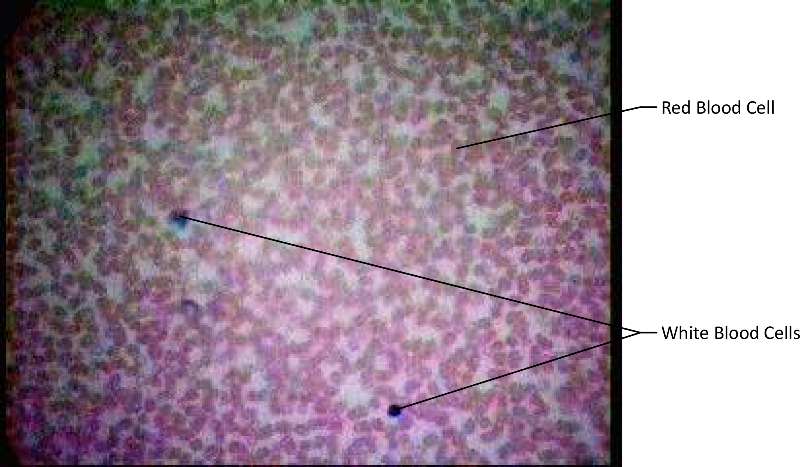
Figure 18. This image shows blood; note the larger size and complexity of white blood cells versus the smaller red disk-shaped red blood cells and is an actual micrograph.
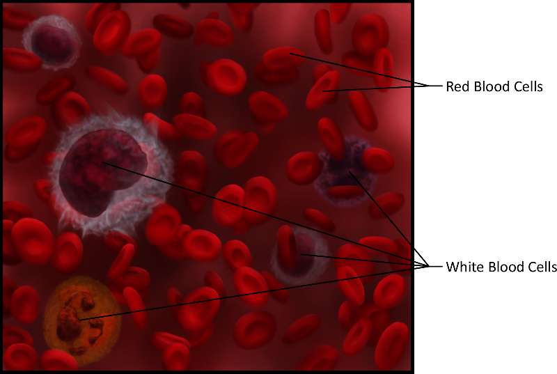
Figure 19. An illustration that shows blood. Notice the larger size and complexity of white blood cells versus the smaller red disk-shaped red blood cells and think about how this relates to their function.
Bone is a connective tissue where osteocytes are located in lacuna in a matrix of calcium salts. Bone functions for support and protection.
The following video discusses bone.
Video 14. View the Bone video on YouTube (opens in a new window)
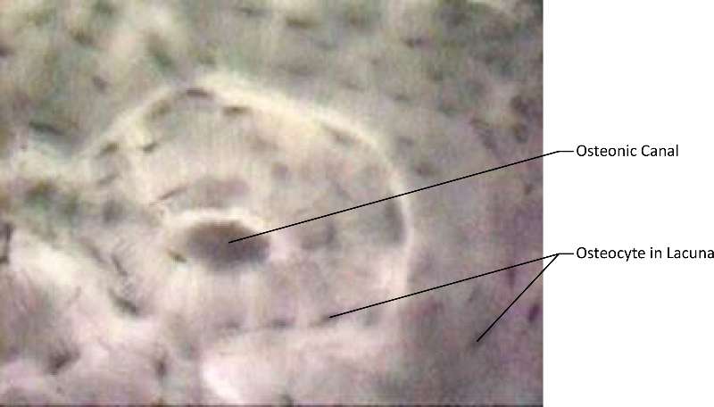
Figure 20. An actual micrograph of bone and that highlights the osteonic canal (which contains the bones blood supplies) and osteocyte(s) in lacuna.
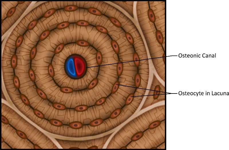
Figure 21. An illustration of bone and highlight the osteonic canal (which contains the bones blood supplies) and osteocyte(s) in lacuna.
Vertebrates have three types of muscle tissue; skeletal, cardiac and smooth. Muscle tissue has the ability to contract or to shorten and thicken to do work. This ability comes from the proteins found inside the muscle cells. The proteins may be arranged so that the cells look striped or striated with alternating light and dark bands. This is the situation with cardiac muscle and skeletal muscle, they are both striated. Smooth muscle is nonstriated because the proteins are arranged differently. Muscle tissue is controlled by the nervous system. Skeletal muscle is controlled by the somatic nervous system which means it is voluntary and under your conscious control. Smooth and cardiac muscle is controlled by the autonomic nervous system which means it is involuntary and you cannot consciously control it.
| Tissue | Structure | Function | Representative Location |
|---|---|---|---|
|
Skeletal Muscle |
polynuclear, long, striated cells |
Move bones |
attached to bones |
|
Smooth Muscle |
small tapered cells with one nucleus centrally located, nonstriated |
peristalsis, constriction of lumen |
walls of hollow organs |
|
Cardiac Muscle |
Uninuclear, striated, branched cells with intercalated disks |
contraction of heart, move blood |
heart |
The following video discusses muscle tissue.
Video 15. View the Muscle Tissue video on YouTube (opens in a new window)
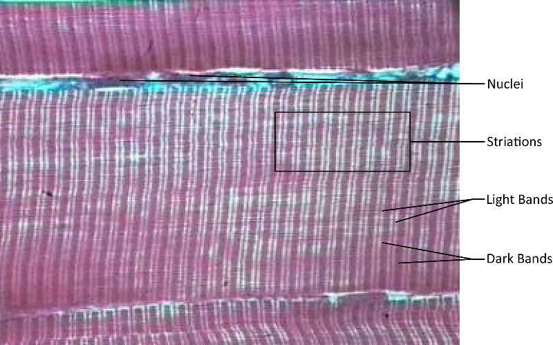
Figure 22. A micrograph of skeletal muscle composed of cell nuclei, striations, light bands and dark bands (dyed purple for ease of visibility).
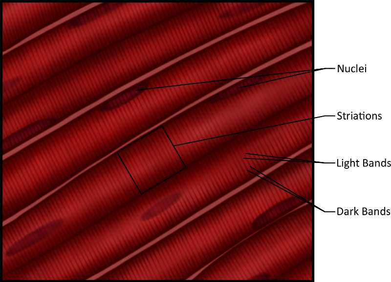
Figure 23. An illustration showing skeletal muscle composed of cell nuclei, striations, light bands and dark bands.
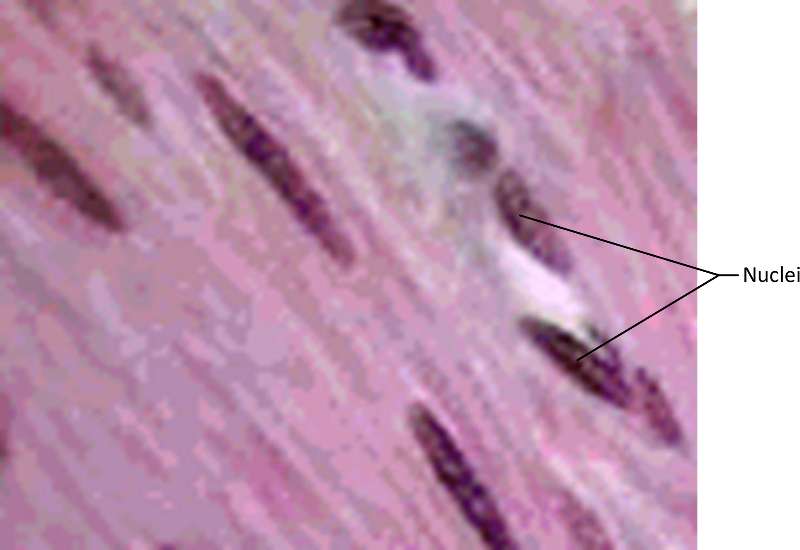
Figure 24. A micrograph showing smooth muscle dyed purple for ease of visibilty The darkest stained cells are the nuclei.
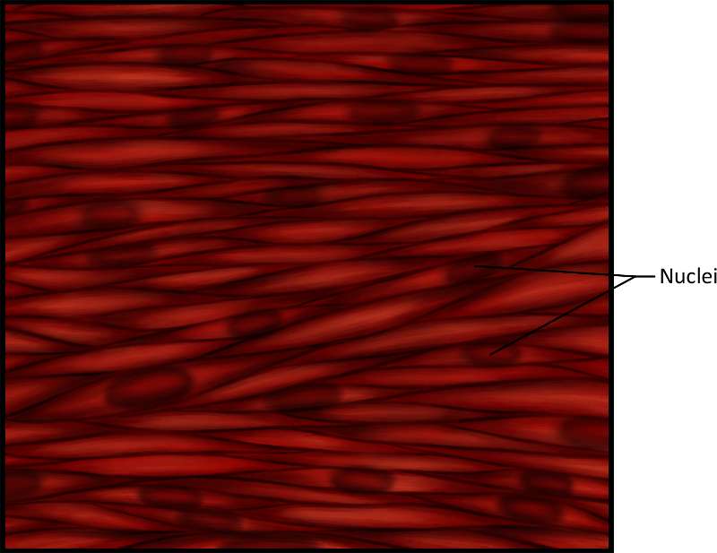
Figure 25. An illustration showing smooth muscle. The darkest stained cellular areas are the nuclei.
 |
|
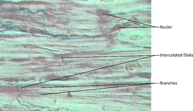
Figure 26. A micrograph (dyed purple for ease of visibilty). Notice the nuclei, branches and intercalated disks (junctions characteristic of cardiac muscle).
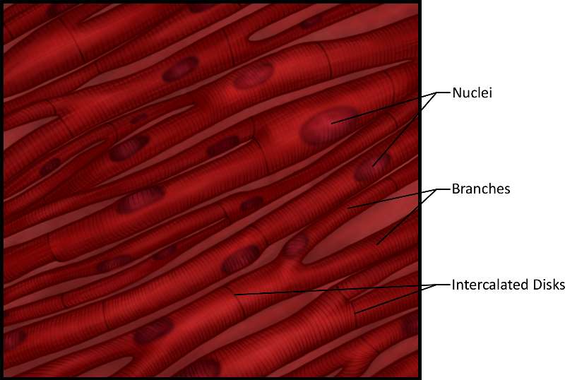
Figure 27. An illustration showing show smooth muscle. Note the nuclei, branches and intercalated disks (junctions characteristic of cardiac muscle).
Nervous Tissue is found in the brain, spinal cord, and nerves. The cell that conducts impulses is called a neuron. The other type of cell is called a neuroglial cell. About 10% of nervous tissue is composed of neurons, the rest of the cells are glial cells. The neurons conduct impulses and the glial cells support the neurons. Students should be able to identify a motor neuron and its parts (axon, dendrite, cell body, synaptic bulb).
The following video discusses nervous tissue.
Video 16. View the Nervous Tissue video on YouTube (opens in new window)
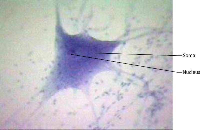
Figure 28. A micrograph showing a motor neuron. Notice the nucleus and soma (cell body) in both.
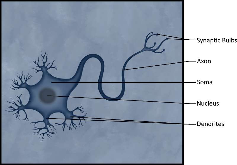
Figure 29. A detailed illustration of a motor neuron showing the axond, dendrites and synaptic bulbs used for transmitting nerve signals.
Organs are composed to two or more tissues working together to accomplish a particular function. Organs, such as membranes, can be as simple as an epithelial tissue and a connective tissue. For example, the conjunctiva that lines your eye lids are composed of simple squamous epithelium and loose connective tissue. Some organs are much more complicated. Consider the small intestine as an example. The small intestine is lined with simple columnar epithelium. The simple columnar epithelium protects the lining but it is also involved with absorption of the nutrients. Beneath the simple columnar epithelium is loose connective tissue. This tissue binds the layers together and is a good medium for the blood vessels that are there to absorb the nutrients and water that have been ingested. There are two layers of smooth muscle in the small intestine. The layer closest to the lumen encircles the lumen. When this layer contracts, the tube constricts. The next layer of smooth muscle runs the length of the intestine. When this longitudinal smooth muscle contracts, the tube shortens. The peristaltic action of the intestine occurs as these muscular layers contract. The small intestine is covered with a membrane composed of simple squamous epithelium and loose connective tissue. As you learn about the different organs in the human body, you will need to think about the tissues they are composed of and how the functions of the tissues allow the organs to perform their functions. Some examples of organs and their different tissues are labeled in the diagrams and micrographs below.
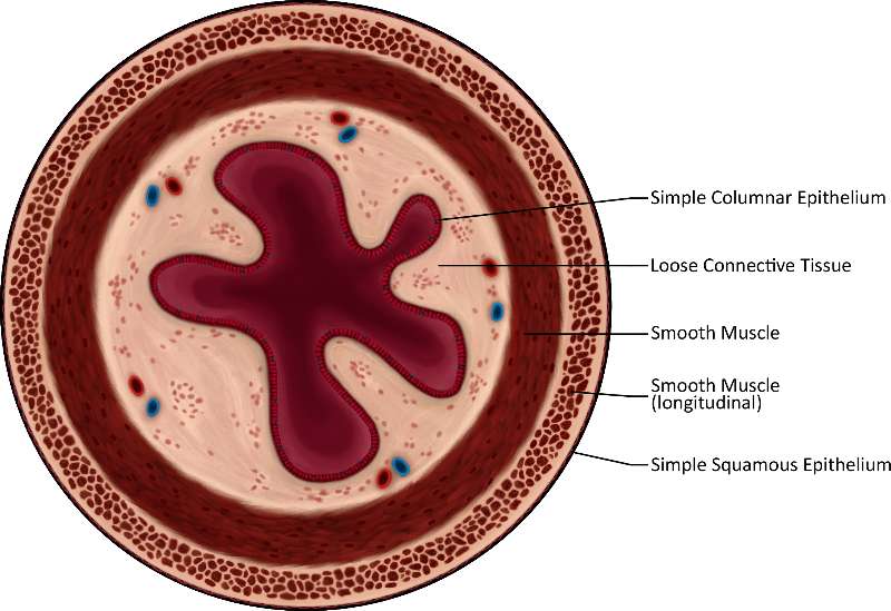
Figure 30. This is a very detailed illustration of the different tissue types fround in a cross-section of the small intestine. The burgundy starburst shape in the center is composed of simple columnar epithelium. The cream colored spongy structure outside of that is composed of loose connective tissue. The burgundy ring around that is composed of smooth muscle and the creme and burgundy very spongy ring outside of that is longitudinal smooth muscle. Lastly, the dark thin ring around the outside of the whole structure is composed of simple squamous epithelium.
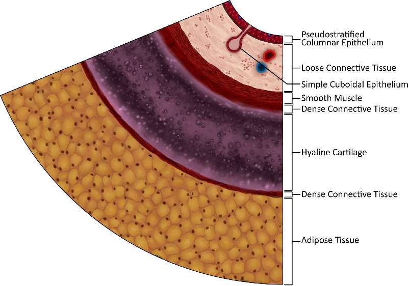
Figure 31. This image is a very detailed illustration of the different tissue types found in a cross-section of the trachea.The layer of the image, closest to this text, is yellow and globular adipose tissue and you can clearly see the fat cell membranes and shaded nuclei. The thick purple layer above that (which seems to contain bubbles) is hyaline cartilage, flanked on either side by thin layers of burgundy dense connective tissue. Above the top layer of dense connective tissue is a layer of burgundy smoth muscle. Atop that is a creme-colored spongy layer of loose connective tissue, intersected by a pink protrusion of simple cuboidal epithelium. Finally, all of these layers are topped by a thin layer of burgundy pseudostratified columnar epithelium.

Except where otherwise noted, this work by The Community College Consortium for Bioscience Credentials is licensed under a Creative Commons Attribution 4.0 International License.;
Text written by Michael V. Ayers, M.S. and Alice Rudolph, M.A. for c3bc.
Instructional Design by Courtney A. Harrington, Ph.D., and Caroline Smith, M.A. for c3bc.
Media by Brittany Clark and Antonio Davis for c3bc.
This product was funded by a grant awarded by the U.S. Department of Labor's Employment and Training Administration. The product was created by the grantee and does not necessarily reflect the official position of the U.S. Department of Labor. The Department of Labor makes no guarantees, warranties, or assurances of any kind, express or implied, with respect to such information, including any information on linked sites and including, but not limited to, accuracy of the information or its completeness, timeliness, usefulness, adequacy, continued availability, or ownership.