Meiotic Cell Division
Anatomy & Physiology I
Another type of cell division, meiosis, is the process used to create gametes. Gametes are the cells used to form offspring. In animals, the gametes are eggs and sperm. In plants, the gametes are ovules and pollen.
In meiosis, just like in mitosis, there is a single duplication event. That is to say, the DNA is doubled only once; in mitosis, this is how the daughter cells contain the right amount of DNA; first it is doubled and then divided in two. Unlike mitosis, which has one division, two divisions occur in meiosis, called Meiosis I and Meiosis II, which results in cells with half as much DNA. As an example, let's imagine chromosomes were apples. If your parent cell has two apples, and they are doubled, you would have four apples, so that in Mitosis when your cell divided, each cell would have 2 apples, the same as the parent. However, in meiosis, you'd split those two cells in half again, so you would have 4 cells with 1 apple each – half the number of apples in the parent cells
So, gametes contain only half of the genetic information found in an organism. That is so the male and female gametes can combine without doubling the amount of genetic material in offspring. For example, in humans, gametes have 23 chromosomes while the rest of our cells have 46 chromosomes (1 set of 23 from our mother's egg and 1 set of 23 from our father's sperm).
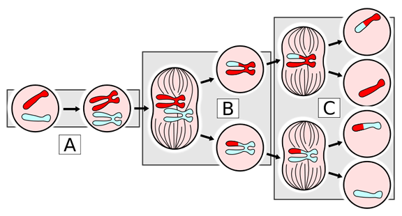
The following figures summarize the Process of Meiosis:
Figure 1. In this figure, the red chromosome represents the maternal while the blue represents the paternal. First, the chromosomes are duplicated, (see A. DNA replication). Then homologous chromosomes separate during Meiosis I, (see B). Then sister chromatid separates in Meiosis II, (see C) giving 4 daughter cells with half the chromosome number of the parent.
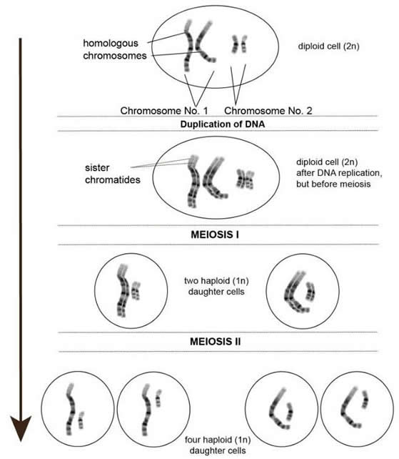
Figure 2. In meiosis, a cell undergoes two divisions: one to separate homologous chromosomes (meiosis I) and one to separate sister chromatids (meiosis II). The result is four haploid daughter cells. In the above diagram, you can see that the chromosomes are duplicated creating sister chromatids in the second picture. After meiosis I you end up with two haploid (1n) daughter cells. In the final picture, meiosis II results in four unique haploid (1n) daughter cells.
Meiosis incorporates two consecutive cycles of division: meiosis I and meiosis II. Before a cell enters meiosis, its DNA is copied, and the duplicated DNA of each chromosome is packed into two sister chromatids. This is the same as what takes place before cell division by mitosis.
Meiosis I and II are each further subdivided into prophase, metaphase, anaphase, and telophase. The first phase of meiosis I is known as prophase I.
During prophase I, the walls of the nucleus begin to break down, homologous chromosomes join together, and, chromosomes crossover and exchange segments. Homologous chromosomes exchange segments during crossing over, which increases the genetic variability in a population. After crossing over occurs, hollow structural tubes, called spindles, spread toward each pole of the cell. These spindles are held in place at the poles of the cell by centrioles, or barrel-shaped rings.
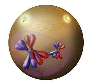
Figure 3. During this phase the nuclear walls begin to break down, homologous chromosomes come together, and chromosomes cross over or exchange genetic material. This is represented by the large blue X lining up with the large red X, and their exchanged genetic material is seen by the mismatched colored tips on each. The same is shown in the smaller blue x and red x, the come together and exchange a small portion of genetic material. After crossing over, spindles form toward each pole shown in a golden yellow and are held in place by the glowing yellow barrel-shaped centrioles.
Next in the process, comes metaphase I. During this phase homologous pairs line up at the center line of the cell. The spindles, which are now completely spread across the cell, bind to each chromosome at its kinetochore.
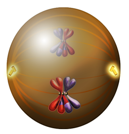
Figure 4. During this phase the homologous chromosomes (large red X and large blue X; small red x and small blue x) line up in the center of the cell and the spindles attach to each at the kinetochore (small grey circle at the center of each X).
During the next stage, anaphase I, the spindles pull one homologue from each pair to opposite poles or sides of the cell.
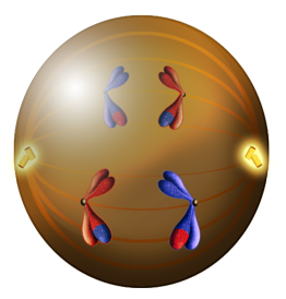
Figure 5. During this phase the spindles pull the homologous chromosomes apart (large blue X and small red x are pulled to the right while the large red X and small blue x are pulled to the left).
In the final phase of meiosis I, telophase I, a nuclear membrane forms around each set of sister chromatids, marking the completion of the first nuclear division. Consequently, the two new cells undergo cytokinesis, a process by which the cytoplasm is divided between the daughter cells.
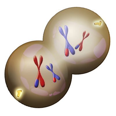
Figure 6. During the final phase of meiosis I, the nuclear membrane begins to reform (shown by the pale whitish circle surrounding each set of chromatids and the cells undergo cytokinesis and cleave the cytoplasm into two new daughter cells.
Match the following Meiosis l phases to the events of each phase.

Almost immediately after cytokinesis in meiosis I, meiosis II commences. In meiosis II, sister chromatids are separated. This division is very similar to a mitotic division and occurs in the same steps as both mitosis and meiosis I.
The key difference between meiosis II and a mitotic division is that crossing over does not occur during meiosis II. When the second meiotic division is completed, four non-identical haploid cells have been produced.
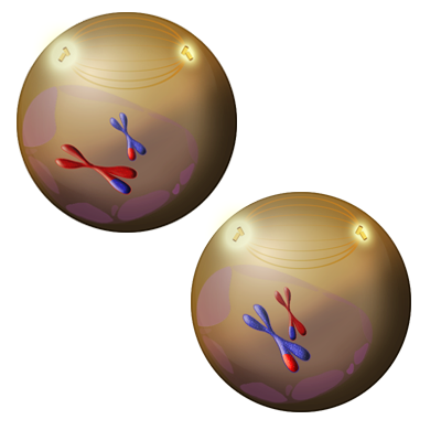
Figure 7. During the first phase of meiosis II, known as Prophase II the nuclear envelope breaks down (shown as a dissipating pale purple circle around the chromatids), and the golden yellow spindles form anchoring themselves at the glowing yellow barrel-shaped centriole.
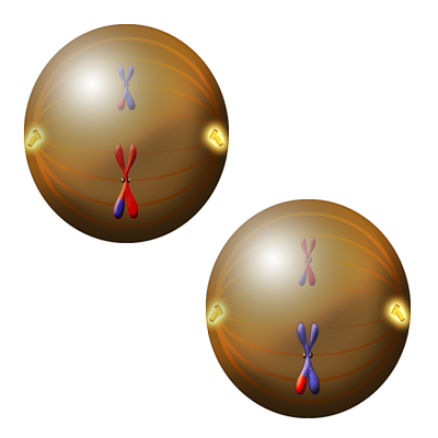
Figure 8. During Metaphase II, the sister chromatids line up across the middle of the cell, and the orange colored spindles attach at the kinetochores (small grey circle at the center of the X).
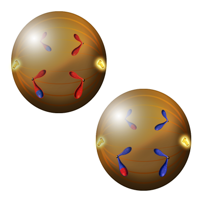
Figure 9. During Anaphase II, the sister chromatids are pulled apart in both cells (looks like each X is being split in half). In the first cell, the small red with a blue tip and large red with a blue tip are pulled to the left while the small red and large red are pulled to the right. In the second cell, the small blue with a red tip and large blue with a red tip are pulled to the left while the small blue and large blue are pulled to the right.
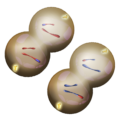
Figure 10. In the final stage of meiosis II, known as Telophase II, the nuclear membrane reforms (shown by the pale whitish circle) around each set of genetic material. The cells then undergo cytokinesis, forming four daughter cells in total.
The following video discusses Meiosis
Video 1: Meiosis (opens YouTube in new window)
Match the following Meiosis ll phases to the events of each phase.
Select the correct image that identifies the different stages of meiosis represented in the activity below.

Select the correct answers for the following questions.

Anaphase I: phase of Meiosis I where the spindles pull one homologue from each pair to opposite poles or sides of the cell.
Anaphase II: phase of Meiosis II when the recombined sister chromatids are pulled apart in both cells (looks like each X is being split in half).
Centrioles: have a role in pulling the duplicated chromosomes to opposite ends of the dividing cell
Crossover: exchange of genetic material between non-sister chromatids resulting in chromosomes that incorporate genes from both parents of the organism
Gametes: specialized cell line that produces germ cells, such as eggs or sperm
Homologous Chromosomes: matched pairs containing the same genes (from both parents) in identical locations along their length.
Meiosis: a nuclear division process that results in four haploid cells
Meiosis I: first round of meiotic cell division; referred to as reduction division because the ploidy level is reduced from diploid to haploid
Meiosis II: second round of meiotic cell division following meiosis I; sister chromatids are separated into individual chromosomes, and the result is four unique haploid cells
Metaphase I: during this phase of Meiosis I, homologous pairs line up at the center line of the cell. The spindles, which are now completely spread across the cell, bind to each chromosome at its kinetochore.
Metaphase II: phase of Meiosis II when the sister chromatids line up across the middle of the cell, and the spindles attach at the kinetochores
Prophase I: phase of Meiosis I when the walls of the nucleus begin to break down, homologous chromosomes join together, and, chromosomes crossover and exchange segments.
Prophase II: phase of Meiosis II when the nuclear envelope breaks down, and the spindles form anchoring themselves at the centriole.
Spindles: hollow structural tubes that assist in chromosome crossover
Telophase I: phase of Meiosis I when a nuclear membrane forms around each set of sister chromatids, marking the completion of the first nuclear division. Consequently, the two new cells undergo cytokinesis, a process by which the cytoplasm is divided between the daughter cells.
Telophase II: phase of Meiosis II when the nuclear membrane reforms around each set of genetic material. The cells then undergo cytokinesis, forming four daughter cells in total.

Except where otherwise noted, this work by The Community College Consortium for Bioscience Credentials is licensed under a Creative Commons Attribution 3.0 Unported License.
Text from BioBook licensed under CC BY NC SA and Boundless Biology Open Textbook licensed under CC BY SA. Modified by Courtney A. Harrington, Ph.D. for c3bc.
Instructional design by Nicole Lipscomb, M.S., Helen Dollyhite, M.A., Caroline Smith, M.A.T., Irene Yee Chief, Ph.D. and Courtney A. Harrington, Ph.D. for c3bc.
Subject Matter Expert: Tiffany Davis and Alice Rudolph, M.A. for c3bc.
Media and interactive objects by Brittany Clark, John Reece and Lucious Oliver, II for c3bc.
This product was funded by a grant awarded by the U.S. Department of Labor's Employment and Training Administration. The product was created by the grantee and does not necessarily reflect the official position of the U.S. Department of Labor. The Department of Labor makes no guarantees, warranties, or assurances of any kind, express or implied, with respect to such information, including any information on linked sites and including, but not limited to, accuracy of the information or its completeness, timeliness, usefulness, adequacy, continued availability, or ownership.