


Extracellular Matrix
Binding of cells in tissues; surface protection, regulation of cellular activities
Intracellular Organization
Anatomy & Physiology I
Close your eyes and picture a brick wall. What is the basic building block of that wall? A single brick, of course. Like a brick wall, your body is composed of basic building blocks, and the building blocks of your body are cells.
Your body has many kinds of cells, each specialized for a specific purpose. Just as a home is made from a variety of building materials, the human body is constructed from many cell types. For example, epithelial cells protect the surface of the body and cover the organs and body cavities within. Bone cells help to support and protect the body. The cells of the immune system fight invading bacteria. Additionally, blood and blood cells carry nutrients and oxygen throughout the body while removing carbon dioxide. Each of these cell types plays a vital role in the growth, development, and day-to-day maintenance of the body. In spite of their enormous variety, however, cells from all organisms—even ones as diverse as bacteria, onion, and human—share certain fundamental characteristics.
Every living organism is made up of cells which are the basic structural and functional unit of all known living organisms. Cells are the smallest units of life. Organisms can be classified as either unicellular or multi-cellular. Unicellular organisms consist of a single cell and include bacteria. Multicellular organisms include plants and animals. Specifically, in multicellular organisms, there are various types of cells, and it is the interactions between these cells that allow organismal functions to occur. The cell, first discovered in 1665 by Robert Hooke, set the foundation for the development of the cell theory, that states that vital functions of an organism occur within cells and that all cells contain the hereditary information.
Now, let us break down the word Eukaryote. The prefix of "eu" means true. We have already learned the definition of "kary" is the nucleus. We can determine that eukaryotes do have a nucleus with this information.
Eukaryotes are typical organisms that include fungi, plants, and animals and are typically larger than prokaryotes. They also contain a nucleus with a double membrane. Eukaryotic cells contain highly structured cytoplasmic structures called organelles and contain one to several thousand mitochondria. These cells are usually multicellular organisms with specialized cells.
Unlike prokaryotic cells, eukaryotic cells have:
Because a membrane surrounds a eukaryotic cell's nucleus, it is often said to have a "true nucleus". The word "organelle" means "little organ", and, as already mentioned, organelles have specialized cellular functions, just as the organs of your body have specialized functions. Organelles allow different functions to be compartmentalized in different areas of the cell.
| Cell Parts | Structure | Function |
|---|---|---|
| Genetic Control | ||
| Nucleus | |
DNA synthesis, RNA synthesis; assembly of ribosomal subunits (in nucleolus) |
| Ribosomes | |
A sphere-shaped structure within the cytoplasm of a cell that is composed of RNA and protein and is the site of protein synthesis. |
| Manufacturing, Distribution, and Breakdown | ||
| Rough Endoplasmic Reticulum | |
Synthesis of membrane lipids and proteins, secretory proteins and hydrolic enzymes; formation of transport vesicles |
| Smooth Endoplasmic Reticulum | |
Synthesis of lipids; detoxification in liver cells; calcium ion storage |
| Golgi Apparatus | |
Modification and sorting of macromolecules formation of lysosomes and transport vesicles |
| Lysosomes | Digestion (food vacuole); storage pf chemicals and cell enlargement (central vacuole); water balance (contractile vacuole) | |
| Peroxisomes | |
Diverse metabolic processes, with breakdown of toxic hydrogen peroxide by-product |
| Vacuoles | |
Contain substances that recently entered the cell, store and transport new molecules |
| Energy Processing | ||
| Mitochondria | Conversion of chemical energy in food to chemical energy of ATP | |
| Structural Support, Movement, and Communication Between Cells | ||
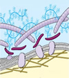
| Cytoskeleton (micro-filaments, intermediate filaments, and microtubules | |
Maintenance of cell shape, anchorage of organelles; movement of organelles within cells; cell movement; bending of cilia and flagella |
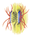
| Cell Junctions | |
Communication between cells; binding of cells in tissues |
|
Extracellular Matrix |
|
Binding of cells in tissues; surface protection, regulation of cellular activities |

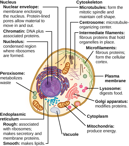
Figure 1. A typical eukaryotic animal cell
CC BY: OpenStax College

Reactive oxygen species (ROS) such as peroxides and free radicals are the highly reactive products of many normal cellular processes, including the mitochondrial reactions that produce ATP and oxygen metabolism. Examples of ROS include the hydroxyl radical OH, H2O2, and superoxide (O2-). Some ROS are important for certain cellular functions, such as cell signaling processes and immune responses against foreign substances. Free radicals are reactive because they contain free unpaired electrons; they can easily oxidize other molecules throughout the cell, causing cellular damage and even cell death. Free radicals are thought to play a role in many destructive processes in the body, from cancer to coronary artery disease.
Peroxisomes, on the other hand, oversee reactions that neutralize free radicals. Peroxisomes produce large amounts of the toxic H2O2 in the process, but peroxisomes contain enzymes that convert H2O2 into water and oxygen. These byproducts are safely released into the cytoplasm. Like miniature sewage treatment plants, peroxisomes neutralize harmful toxins so that they do not wreak havoc in the cells. The liver is the organ primarily responsible for detoxifying the blood before it travels throughout the body, and liver cells contain an exceptionally high number of peroxisomes.
Defense mechanisms such as detoxification within the peroxisome and certain cellular antioxidants serve to neutralize many of these molecules. Some vitamins and other substances, found primarily in fruits and vegetables, have antioxidant properties. Antioxidants work by being oxidized themselves, halting the destructive reaction.
Oxidative stress is the term used to describe damage to cellular components caused by ROS. Due to their characteristic unpaired electrons, ROS can set off chain reactions where they remove electrons from other molecules, which then become oxidized and reactive, and do the same to other molecules, causing a chain reaction. ROS can cause permanent damage to cellular lipids, proteins, carbohydrates, and nucleic acids. Damaged DNA can lead to genetic mutations and even cancer. A mutation is a change in the nucleotide sequence in a gene within a cell's DNA, potentially altering the protein coded by that gene. Other diseases believed to be triggered or exacerbated by ROS include Alzheimer's disease, cardiovascular diseases, diabetes, Parkinson's disease, arthritis, Huntington's disease, and schizophrenia, among many others. It is noteworthy that these diseases are largely age-related. Many scientists believe that oxidative stress is a major contributor to the aging process.
The Theory of Free Radicals within Cells
The free radical theory on aging was originally proposed in the 1950s, and still remains under debate. Generally speaking, the free radical theory of aging suggests that accumulated cellular damage from oxidative stress contributes to the physiological and anatomical effects of aging. There are two significantly different versions of this theory: one states that the aging process itself is a result of oxidative damage, and the other states that oxidative damage causes age-related disease and disorders. The latter version of the theory is more widely accepted than the former. However, many lines of evidence suggest that oxidative damage does contribute to the aging process. Research has shown that reducing oxidative damage can result in a longer lifespan in certain organisms such as yeast, worms, and fruit flies. Conversely, increasing oxidative damage can shorten the lifespan of mice and worms. Interestingly, a manipulation called calorie restriction (moderately restricting the caloric intake) has been shown to increase life span in some laboratory animals.
It is believed that this increase is at least in part due to a reduction of oxidative stress. However, a long-term study of primates with calorie-restriction showed no increase in their lifespan. A great deal of additional research will be required to better understand the link between reactive oxygen species and aging.
The following video introduces DNA:
Video 1. Click this link to view Overview of DNA on YouTube (opens in a new window).
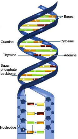
Figure 2. DNA is shaped like a double helix, which resembles a twisted ladder. There are two strands of DNA with the complimentary nucleotides from each pointing inward from each strand. The sugar-phosphate backbone is shown in blue while the nucleotides are shown as follows: guanine "G" is shown in green that pairs with cytosine "C" shown in orange. Adenine "A" is shown in yellow and pairs with thymine "T" shown in red.
Genes are the discrete units of heredity contained within the DNA double helices of chromosomes. A gene is a specific stretch of DNA and RNA that codes for a type of protein, or for an RNA chain, which has a function in the organism. Living things depend on genes because they specify all proteins and functional RNA chains.
So why are genes important? Genes hold the information to build and maintain an organism's cells and pass genetic traits to offspring. All organisms have many genes corresponding to various biological traits. Some of these traits are immediately visible, such as eye color or number of limbs. However, some of these traits are not immediately apparent, such as blood type, increased risk for specific diseases, or the thousands of basic biochemical processes that comprise life.
The following video explains how DNA is structured:
Video 2. Click this link to view Structure of DNA on YouTube (opens in new window).
As we discussed in the macromolecules module, DNA consists of two long polymers of simple units called nucleotides.
Each nucleotide has three components:
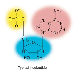
Figure 3. In the diagram above, the phosphate is highlighted in yellow, the sugar (in this case ribose) in blue, and the nitrogenous base in pink.
The nucleotides are arranged so that the DNA strands have backbones made of sugars and phosphate groups joined by ester bonds. The backbone of DNA forms the outside of each of the two helices, with the nucleotide bases bound together and stacked in the center of the helix.
The nitrogenous bases found in DNA are:
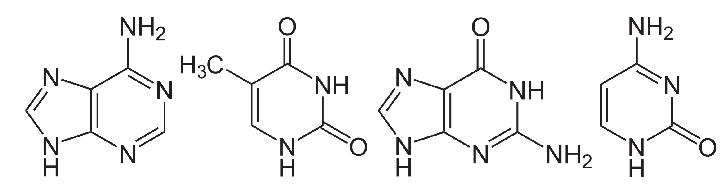
Figure 4. This diagram shows the chemical structure of the nitrogenous bases found in DNA (from left to right): adenine (A), thymine (T), guanine (G), and cytosine (C).
Since Watson and Crick first published the DNA structure, we have learned that DNA replication, or the copying of DNA, is a biological process that occurs in all living organisms and is the basis for biological inheritance. The process starts with one double-stranded DNA molecule and produces two identical copies of the molecule. Each strand of the original double-stranded DNA molecule serves as a template for the production of the complementary strand. Cellular proofreading and error correcting mechanisms ensure a near perfect result for DNA replication.
Test your knowledge by completing the quiz below.

During DNA replication, each of the two strands of DNA serves as the template for the synthesis of a new, complementary strand.
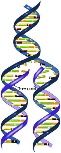
Figure 5. This image represents a simplified model of DNA Replication. At the top, the parental DNA contains two strands of DNA with hydrogen bonds between complimentary bases. In the middle of the figure, the two parental DNA strands begin to separate in the first step of replication, leaving both strands available to act as templates for new DNA. At the bottom of the figure, complementary base pairs align with the templates and connect at the sugar-phosphate backbone to form new DNA molecules, each with a single old and new strand.
Watson and Crick hypothesized that DNA replication occurred when the double helix separated, and the two strands acted as templates for new strand synthesis, resulting in two double strands that each contains one old and one newly replicated strand. The model of DNA replication that Watson and Crick proposed is called the semiconservative model because it results in two daughter double helices that each contains one old strand and one newly replicated strand of DNA.
The following video explains DNA replication:
Video 3. Click this link to view DNA Replication - What is it? on YouTube (opens in new window).
In eukaryotes, DNA replication is controlled within the cell cycle as we discussed in the beginning of Unit 4. As the cell grows and divides, it progresses through stages. DNA replication occurs during the synthesis, or S phase. Most bacteria, or prokaryotes, do not go through a well-defined cell cycle, but instead continuously copy their DNA. During rapid growth, this can result in the concurrent occurrences of multiple rounds of replication.
DNA replication begins with the unzipping of the parent molecule as the hydrogen bonds between the base pairs are broken. The process is initiated at particular points in the DNA, known as origins, which are targeted by proteins that separate the two strands and initiate DNA synthesis. Origins of replication contain DNA sequences recognized by replication initiator proteins. These initiator proteins recruit other proteins to separate the two strands at the origin, forming a bubble. As DNA synthesis continues, the original DNA strands unwind on each side of the bubble, forming a replication fork with two prongs at each end of the bubble.
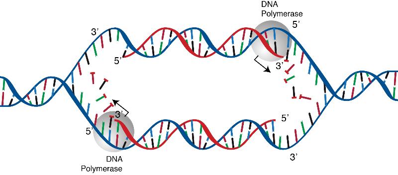
Figure 6. Summary of DNA Replication. In DNA Replication, the bonds are broken between the two strands of the DNA double helix, then the DNA is then unwound, and both sides of the double helix used as templates to build new strands of complementary DNA. The result is two new copies of the DNA, each one having an old and a new strand.
Test your knowledge by completing the quiz below.
Transcription Regulation constitutes a major component of biological activities in the cell. A key function of transcriptional activity is the control of gene expression necessary for the maintenance of a cellular state. In metazoans, all cell types, except mature germ cells, contain the same genetic information within the species. Yet, each cell type expresses a unique subset of all genes encoded. This is because each type of cell expresses different genes, a mechanism known as "differential gene expression."
Differential gene expression can be controlled at many levels. Transcription occurs at the first stage of expression. Coding and non-coding genes contain promoter and enhancer sequences that are bound by transcription activators and repressors, known as transcription factors (TFs). The levels and activities of TF's determine the extent of transcription.
Regulation of gene expression refers to the control of the amount and the timing of appearance of the functional product of a gene. Control of gene expression is vital to allow a cell to produce the gene products it needs when it needs them; this, in turn, provides cells with the flexibility to adapt to a variable environment, external signals, damage to the cell, etc. Gene expression in organisms is carefully regulated to allow them to adapt to variable conditions and, most importantly, to prevent wasteful overproduction of unneeded proteins, which would make the organism not productive.
Organisms regulate expression of their genes to achieve developmental changes, cellular specialization, and adaptation to a new environment. The expression of genetic information in a given cell or organism is neither random nor fully pre-programmed. The information in an organism's genome must be regulated in an orderly fashion during development and, yet, must be available to direct the organism's responses to changes in internal and external conditions. Thus, every step of the pathway, from gene expression to protein synthesis, localization, and degradation, can be controlled to maximize the cell's survivability and efficiency.
Cell Junctions: Communication between cells; binding of cells in tissues.
Cytoskeleton: Maintenance of cell shape, anchorage of organelles; movement of organelles within cells; cell movement; bending of cilia and flagella.
Differential Gene Expression: Active interpretation of the information coded in a gene to produce a functional gene productl; in this case, it can be controlled at many levels.
Eukaryotes: Typical organisms that include fungi, plants, and animals and are typically larger than prokaryotes. They also contain a nucleus with a double membrane.
Extracellular Matrix: Binding of cells in tissues; surface protection, regulation of cellular activities.
Golgi Apparatus: Modification and sorting of macromolecules formation of lysosomes and transport vesicles.
Lysosomes: Digestion (food vacuole); storage of chemicals and cell enlargement (central vacuole); water balance (contractile vacuole).
Mitochondria: Conversion of chemical energy in food to chemical energy of ATP.
Mutation: a change in the nucleotide sequence in a gene within a cell's DNA, potentially altering the protein coded by that gene.
Nucleus: DNA synthesis, RNA synthesis; assembly of ribosomal subunits (in nucleolus).
Oxidative Stress: the term used to describe damage to cellular components caused by ROS.
Peroxisomes: Diverse metabolic processes, with breakdown of toxic hydrogen peroxide by-product.
Reactive Oxygen Species (ROS): such as peroxides and free radicals are the highly reactive products of many normal cellular processes, including the mitochondrial reactions that produce ATP and oxygen metabolism.
Ribosomes: A sphere-shaped structure within the cytoplasm of a cell that is composed of RNA and protein and is the site of protein synthesis.
Rough Endoplasmic Reticulum: Synthesis of membrane lipids and proteins, secretory proteins and hydraulic enzymes; formation of transport vesicles.
Smooth Endoplasmic Reticulum: Synthesis of lipids; detoxification in liver cells; calcium ion storage.
Transcription Regulation: the control of the transcription component of gene expression necessary for the maintenance of a cellular state.
Vacuoles: Contain substances that recently entered the cell, store and transport new molecules.

Except where otherwise noted, this work by The Community College Consortium for Bioscience Credentials is licensed under a Creative Commons Attribution 3.0 Unported License.
Text from BioBook licensed under CC BY NC SA and Boundless Biology Open Textbook licensed under CC BY SA. Modified by Courtney A. Harrington, Ph.D. for c3bc.
Instructional design by Nicole Lipscomb, M.S., Helen Dollyhite, M.A., Irene Yee Chief, Ph.D., Caroline Smith, M.A, and Courtney A. Harrington, Ph.D. for c3bc.
Subject Matter Expert: Tiffany Davis for c3bc.
Reviewed by Shelton M. Charles, Ph.D and Alice Rudolph, M.A. for c3bc.
Media and Interactive objects by Joe deCastro, John Reece, Brittany Clark and Lucious Oliver II for c3bc.
This product was funded by a grant awarded by the U.S. Department of Labor's Employment and Training Administration. The product was created by the grantee and does not necessarily reflect the official position of the U.S. Department of Labor. The Department of Labor makes no guarantees, warranties, or assurances of any kind, express or implied, with respect to such information, including any information on linked sites and including, but not limited to, accuracy of the information or its completeness, timeliness, usefulness, adequacy, continued availability, or ownership.