Anatomical Terminology
Anatomy & Physiology I
Introduction to Anatomy and Physiology
Objectives
- Distinguish between anatomy and physiology.
- Identify the functional characteristics of human life.
- Use appropriate anatomical terminology to identify key body structures, regions, and directions in the body.
- Compare and contrast at least four medical imagining techniques
Though you may approach a course in anatomy and physiology strictly focused on your field of study, the knowledge that you gain in this course will serve you well in many aspects of your life. An understanding of anatomy and physiology is not only fundamental to any health profession, but it can also benefit your own health.
Familiarity with the human body can help you make healthful choices and prompt you to take appropriate action when signs of illness arise. Your knowledge in this field will help you understand news about nutrition, medications, medical devices, and procedures; as well as, help you understand genetic or infectious diseases. At some point, everyone will have a problem with some aspect of his or her body, so your knowledge will help you to be a better parent, spouse, partner, friend, colleague, or caregiver.
Anatomy
Human anatomy is the study of the structures of the body. The word anatomy comes from a Greek root meaning to cut apart. Human anatomy was first studied by observing the exterior of the body and observing the wounds and injuries of soldiers. Later, physicians were allowed to dissect dead bodies in order to augment their knowledge. When a body is dissected, its structures are cut apart in order to observe their physical attributes and their relationships to one another. Dissection is still used in medical schools, anatomy courses, and in pathology labs. In order to observe structures in living people, however, a number of imaging techniques have been developed. These techniques allow clinicians to visualize structures inside the living body such as a cancerous tumor or a fractured bone.
Anatomical Specializations
Like most scientific disciplines, anatomy has areas of specialization. Gross anatomy is the study of the larger structures of the body, those visible without the aid of magnification (Figure 1a). Macro- means large, thus, gross anatomy is also referred to as macroscopic anatomy. In contrast, micro- means small, and microscopic anatomy is the study of structures that can be observed only with the use of a microscope or other magnification devices (Figure 1b). Microscopic anatomy includes cytology, the study of cells and histology, the study of tissues. As the technology of microscopes has advanced, anatomists have been able to observe smaller and smaller structures of the body, from slices of large structures like the heart, to the three-dimensional structures of large molecules in the body.
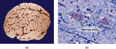
Figure 1. Gross and Microscopic Anatomy
(a) Gross anatomy considers large structures such as the brain. (b) Microscopic anatomy can deal with the same structures, though at a different scale. This is a micrograph of nerve cells from the brain. Light Microscopy × 1600.
(Credit a: "WriterHound"/Wikimedia Commons; credit b: Micrograph provided by the Regents of University of Michigan Medical School c 2012)
CC By: OpenStax College
Anatomical Approaches
Anatomy has two approaches. Regional anatomy helps us appreciate the interrelationships of body structures, such as how muscles, nerves, blood vessels, and other structures work together to serve a particular body region. Systemic anatomy is the study of the structures that make up a discrete body system or group of structures that work together to perform a unique body function. For example, a systemic anatomical study of the muscular system would consider all of the skeletal muscles of the body. This course will utilize a systemic approach to anatomy.
Physiology
While anatomy is all about structure, physiology is about function. Human physiology is the scientific study of the chemistry and physics of the structures of the body, and the ways in which they work together to support the functions of life. Much of the study of physiology centers on the body's tendency toward homeostasis. Homeostasis is the state of steady internal conditions maintained by living things and you will learn much more about this in the next module. The study of physiology certainly includes observation, both with the naked eye and with microscopes, as well as manipulations and measurements. However, current advances in physiology usually depend on carefully designed laboratory experiments that reveal the functions of the many structures and chemical compounds that make up the human body.
Physiological Specializations
Like anatomists, physiologists typically specialize in a particular branch of physiology. For example, neurophysiology is the study of the brain, spinal cord, and nerves and how these work together to perform functions as complex and diverse as vision, movement, and thinking. Physiologists may work from the organ level (i. e. exploring what different parts of the brain do), all the way to the molecular level (i. e. exploring how an electrochemical signal travels along nerves).
Anatomy and Physiology Interrelationships
Structure and function of the human body are closely related. The structure of a bone or muscle or organ is such that it can do specific functions. Function will change if the structure of an organ, or even the chemicals of the body, change. For instance, bones of the skull protect the brain, thin air sacs of the lungs permit movement of gases across them, and the durable keratin protein of your nails protects the frequently used fingers. Even the three-dimensional structure of certain molecules is essential to their function.
Take a minute to come up with other examples of how the structure of skin allows for its function. What about the connection between the location of the heart and its function? The study of A & P will make much more sense if you will look for a structure's function, as you study it. Ask yourself, "Why is this shaped as such? Why is it located here? And What is its use?"

What is Life?
I think that this is a very important question to revisit when we study concepts as critical to human life as anatomy and physiology. Defining life is not a simple thing to do; this is quite evident in many of the societal issues that make the headlines so often today. For example, when does one actually die?
People can be kept alive for years on life support, despite being brain dead. Also, when one's heart is no longer working, it may still be possible to survive, with the help of a machine, until a donor heart can be found. Are there medical scenarios in which it is ok to pull that plug? How about the abortion issue: when is abortion ok?
The point I want make is that science can not and should not answer these questions. Science can give us knowledge that will be useful in making our personal decisions. However, an individual's feelings about these issues involves mixing scientific knowledge with their own moral values. Science can not make moral or religious judgments; however, before anyone reaches a conclusion about a major issue, it's definitely important to gather as much knowledge about the issue as possible. It is also important to evaluate the source of the information for potential bias.
The following video discusses life as a controversial topic.
Video 1. View the Controversial Topic video on YouTube (opens in new window).
The following video discusses universal properites of life.
Video 2. View the What is Life? video on YouTube (opens in new window).
The Seven Properties of Life
There are common characteristics shared by all living things. These characteristics define whether something can be considered alive, and therefore are included when we study the human body. These characteristics link such different organisms as bacteria and humans.
The seven properties of life are as follows:
- Order: Living things display an ordered organization.
- Organisms are highly organized, coordinated structures that consist of one or more cells. Even very simple, single-celled organisms are remarkably complex: inside each cell, atoms make up molecules; these in turn make up cell organelles and other cellular inclusions. In multicellular organisms, similar cells form tissues. Tissues, in turn, collaborate to create organs (body structures with a distinct function). Organs work together to form organ systems.
- Reproduction: Living things reproduce more of their own kind.
- For example, these adult humans produced twin babies.

Figure 2. Adult humans with twin babies
CC BY: Xayana
- Regulation: Living things have systems in place to maintain a constant internal environment despite fluctuating external conditions. This property of life is sometimes also called homeostasis.
- Even the smallest organisms are complex and require multiple regulatory mechanisms to coordinate internal functions, respond to stimuli, and cope with environmental stresses. Two examples of internal functions regulated in an organism are nutrient transport and blood flow. Organs (groups of tissues working together) perform specific functions, such as carrying oxygen throughout the body, removing wastes, delivering nutrients to every cell, and cooling the body.
- Energy Processing: Living things take in energy and convert it to useful forms in order to perform work.
- All organisms use a source of energy for their metabolic activities. Some organisms capture energy from the sun and convert it into chemical energy in food; others use chemical energy in molecules they take in as food.
- Growth and Development: Things that are alive grow and develop.
- All organisms grow and develop following specific instructions coded for by their genes. These genes provide instructions that will direct cellular growth and development, ensuring that a species' young will grow up to exhibit many of the same characteristics as its parents.
- Response to the Environment: Living things respond to stimuli.
- Organisms respond to diverse stimuli. For example, when we put a hand on a hot stove, we respond by contracting the biceps muscle in the arm. Movement toward a stimulus is considered a positive response, while movement away from a stimulus is considered a negative response.
- Evolutionary Adaptation: Populations of living things evolve over time in order to adapt to their environment.
- For example, humans (and other mammals) have evolved arrector pili muscles attached to body hairs, making them stand on end, for insulation and/or a fear response.
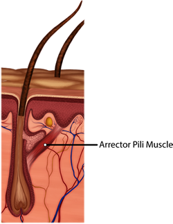
Figure 3. Arrector pili muscles (highlighted in orange) cause mammal hairs to stand up, when cold and/or afraid.

Anatomical Position
The purpose of anatomical terminology is not to confuse, but rather to increase precision and reduce medical errors.These terms derive from ancient Greek and Latin words. Because these languages are no longer used in everyday conversation, the meaning of their words does not change.To further increase precision, anatomists standardize the way in which they view the body. Just as maps are normally oriented with north at the top, the standard body "map," or anatomical position, is that of the body standing upright, with the feet at shoulder width and parallel, toes forward. The upper limbs are held out to each side, and the palms of the hands face forward as illustrated in Figure 4. Using this standard position reduces confusion. It does not matter how the body being described is oriented, the terms are used as if it is in anatomical position. For example, a scar in the "anterior (front) carpal (wrist) region" would be present on the palm side of the wrist. The term "anterior" would be used even if the hand were palm down on a table.
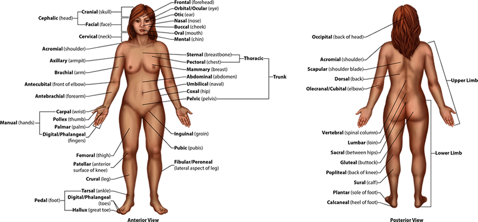
Figure 4. Regions of the Human Body. The human body is shown in anatomical position in an anterior view and a posterior view. The regions of the body are labeled in boldface.
A body that is lying down is described as either prone or supine. Prone describes a face-down orientation, and supine describes a face up orientation. These terms are sometimes used in describing the position of the body during specific physical examinations or surgical procedures.
Instructions: Drag the lable from the bottom to the correct anatomical position.

Regional Terms
The human body's numerous regions have specific terms to help increase precision (see Figure 4). Notice that the term "brachium" or "arm" is reserved for the "upper arm" and "antebrachium" or "forearm" is used rather than "lower arm." Similarly, "femur" or "thigh" is correct, and "leg" or "crus" is reserved for the portion of the lower limb between the knee and the ankle. You will be able to describe the body's regions using the terms from the figure.
Directional Terms
Certain directional anatomical terms appear throughout this and any other anatomy textbook (Figure 5). These terms are essential for describing the relative locations of different body structures. For instance, an anatomist might describe one band of tissue as "inferior to" another or a physician might describe a tumor as "superficial to" a deeper body structure. Commit these terms to memory to avoid confusion when you are studying or describing the locations of particular body parts.
- Anterior (or ventral) describes the front or direction toward the front of the body. The toes are anterior to the foot.
- Posterior (or dorsal) describes the back or direction toward the back of the body. The popliteus is posterior to the patella.
- Superior (or cranial) describes a position above or higher than another part of the body proper. The orbits are superior to the oris.
- Inferior (or caudal) describes a position below or lower than another part of the body proper; near or toward the tail (in humans, the coccyx, or lowest part of the spinal column). The pelvis is inferior to the abdomen.
- Lateral describes the side or direction toward the side of the body. The thumb (pollex) is lateral to the digits.
- Medial describes the middle or direction toward the middle of the body. The hallux is the medial toe.
- Proximal describes a position in a limb that is nearer to the point of attachment or the trunk of the body. The brachium is proximal to the antebrachium.
- Distal describes a position in a limb that is farther from the point of attachment or the trunk of the body. The crus (crural) is distal to the femur (femoral).
- Superficial describes a position closer to the surface of the body. The skin is superficial to the bones.
- Deep describes a position farther from the surface of the body. The brain is deep to the skull.

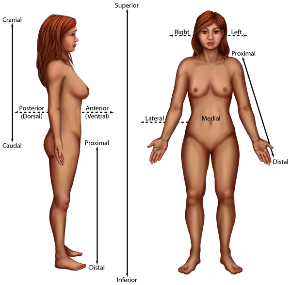
Figure 5. Directional Terms Applied to the Human Body. Paired directional terms are shown as applied to the human body.
CC BY: OpenStax College
Instructions: Drag the lable from the bottom to the correct anatomical position.
Body Planes
A section is a two-dimensional surface of a three-dimensional structure that has been cut. Modern medical imaging devices enable clinicians to obtain "virtual sections" of living bodies. We call these scans. Body sections and scans can be correctly interpreted, however, only if the viewer understands the plane along which the section was made. A plane is an imaginary two-dimensional surface that passes through the body. There are three planes commonly referred to in anatomy and medicine, as illustrated in Figure 6.
- The sagittal plane is the plane that divides the body or an organ vertically into right and left sides. If this vertical plane runs directly down the middle of the body, it is called the midsagittal or median plane. If it divides the body into unequal right and left sides, it is called a parasagittal plane or less commonly a longitudinal section.
- The frontal plane is the plane that divides the body or an organ into an anterior (front) portion and a posterior (rear) portion. The frontal plane is often referred to as a coronal plane. ("Corona" is Latin for "crown.")
- The transverse plane is the plane that divides the body into superior and inferior parts. Transverse planes produce images referred to as cross sections.
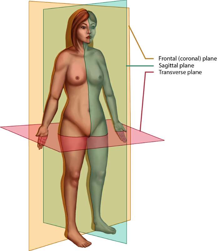
Figure 6. The three planes of the body most commonly used in anatomical and medical imaging are the sagittal, frontal (or coronal), and transverse plane.
The following video illustrates the orientation of different anatomical body planes on an apple.
Video 3. View the Planes & Sections video on YouTube (opens in new window).

Medical Imaging
Early X-Rays
German physicist Wilhelm Röntgen (1845–1923) was experimenting with electrical current when he discovered that a mysterious and invisible "ray" would pass through his flesh but leave an outline of his bones on a screen coated with a metal compound. In 1895, Röntgen made the first durable record of the internal parts of a living human: an "X-ray" image of his wife's hand. In 1901, Röntgen was awarded the first Nobel Prize for physics for his work in this field.
The X-ray is a form of high energy electromagnetic radiation with a short wavelength capable of penetrating solids and ionizing gases. As they are used in medicine, X-rays are emitted from an X-ray machine and directed toward a specially treated metallic plate placed behind the patient's body. The beam of radiation results in darkening of the X-ray plate. X-rays are slightly impeded by soft tissues, which show up as gray on the X-ray plate, whereas hard tissues, such as bone, largely block the rays, producing a light-toned "shadow" in the two-dimensional film image. Thus, X-rays are best used to visualize hard body structures such as teeth and bones. Like many forms of high energy radiation, however, X-rays are capable of damaging cells and initiating changes that can lead to cancer. This danger of excessive exposure to X-rays was not fully appreciated for many years after their widespread use.The disadvantage of radiation to the patient and the operator is now addressed with proper shielding and by limiting exposure.
Computed Tomography
Tomography refers to imaging by sections. Computed tomography (CT) is a noninvasive imaging technique that uses computers to analyze several cross-sectional X-rays in order to reveal minute details about structures in the body (Figure 7a).The technique was invented in the 1970s and is based on the principle that, as X-rays pass through the body, they are absorbed or reflected at different levels. In the technique, a patient lies on a motorized platform while a computerized axial tomography (CAT) scanner rotates 360 degrees around the patient, taking X-ray images. A computer combines these images into a two-dimensional view of the scanned area, or "slice."
Since 1970, the development of more powerful computers and more sophisticated software has made CT scanning routine for many types of diagnostic evaluations. It is especially useful for soft tissue scanning, such as of the brain. Its level of detail is so precise that it can allow physicians to measure the size of a mass down to a millimeter. The main disadvantage of CT scanning is that it exposes patients to a dose of radiation many times higher than that of X-rays. In fact, children who undergo CT scans are at increased risk of developing cancer, as are adults who have multiple CT scans.
Magnetic Resonance Imaging
Magnetic resonance imaging (MRI) is a noninvasive medical imaging technique based on a phenomenon of nuclear physics discovered in the 1930s, in which matter exposed to magnetic fields and radio waves was found to emit radio signals. In 1970, a physician and researcher named Raymond Damadian noticed that malignant (cancerous) tissue gave off different signals than normal body tissue. He applied for a patent for the first MRI scanning device, which was in use clinically by the early 1980s. The early MRI scanners were crude, but advances in digital computing and electronics led to their advancement over any other technique for precise imaging, especially to discover tumors. MRI also has the major advantage of not exposing patients to radiation.
Drawbacks of MRI scans include their much higher cost, and patient discomfort with the procedure. The MRI scanner subjects the patient to such powerful electromagnets that the scan room must be shielded. The patient must be enclosed in a metal tube-like device for the duration of the scan (see Figure 7b), sometimes as long as thirty minutes, which can be impractical for ill patients. The device is also so noisy that, even with earplugs, patients can become anxious or even fearful. These problems have been overcome somewhat with the development of "open" MRI scanning, which does not require the patient to be entirely enclosed in the metal tube. Patients with iron-containing metallic implants cannot undergo MRI scanning because it can dislodge these implants.
Positron Emission Tomography
Positron emission tomography (PET) is a medical imaging technique involving the use of so-called radiopharmaceuticals, substances that emit radiation that is short-lived and therefore relatively safe to administer to the body. Although the first PET scanner was introduced in 1961, it took 15 more years before radiopharmaceuticals were combined with the technique and revolutionized its potential. The main advantage is that PET (see Figure 7c) can illustrate physiologic activity, including nutrient metabolism and blood flow, of the organ or organs being targeted. While CT and MRI scans can only show static images. PET is widely used to diagnose a multitude of conditions, such as heart disease, the spread of cancer, certain forms of infection, brain abnormalities, bone disease, and thyroid disease.
Ultrasonography
Ultrasonography is an imaging technique that uses the transmission of high-frequency sound waves into the body to generate an echo signal that is converted by a computer into a real-time image of anatomy and physiology (see Figure 7d). Ultrasonography is the least invasive of all imaging techniques, and it is therefore used more freely in sensitive situations such as pregnancy. The technology was first developed in the 1940s and 1950s. Ultrasonography is used to study heart function, blood flow in the neck or extremities, certain conditions such as gallbladder disease, and fetal growth and development. The main disadvantages of ultrasonography are that the image quality is heavily operator-dependent and that it is unable to penetrate bone and gas.
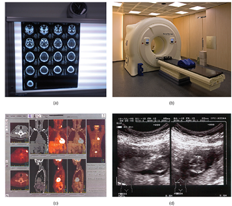
Figure 7. Medical Imaging Techniques. (a) The results of a CT scan of the head are shown as successive transverse sections. (b) An MRI machine generates a magnetic field around a patient. (c) PET scans use radiopharmaceuticals to create images of active blood flow and physiologic activity of the organ or organs being targeted. (d) Ultrasound technology is used to monitor pregnancies because it is the least invasive of imaging techniques and uses no electromagnetic radiation. (Credit a: Akira Ohgaki/flickr; credit b: "Digital Cate"/flickr; credit c: "Raziel"/Wikimedia Commons; credit d: "Isis"/Wikimedia Commons)
CC BY: OpenStax College
Self Assessment
Glossary
Anatomical Position: the body standing upright, with the feet at shoulder width and parallel, toes forward.
Anatomy: science that studies the form and composition of the body's structures.
Anterior: (or ventral) describes the front or direction toward the front of the body.
Computed Tomography (CT): a noninvasive imaging technique that uses computers to analyze several cross-sectional X-rays in order to reveal minute details about structures in the body
Cytology: the study of cells.
Deep: describes a position farther from the surface of the body.
Distal: describes a position in a limb that is farther from the point of attachment or the trunk of the body.
Energy Processing: Living things take in energy and convert it to useful forms in order to perform work.
Evolutionary Adaptation: Populations of living things evolve over time in order to adapt to their environment.
Frontal plane: (or coronal) is the plane that divides the body or an organ into an anterior (front) portion and a posterior (rear) portion.
Gross Anatomy: study of the larger structures of the body, typically with the unaided eye; also referred to macroscopic anatomy.
Growth and Development: Things that are alive grow and develop.
Histology: the study of tissues.
Inferior: (or caudal) describes a position below or lower than another part of the body proper; near or toward the tail (in humans, the coccyx, or lowest part of the spinal column).
Lateral: describes the side or direction toward the side of the body.
Magnetic resonance imaging (MRI): a noninvasive medical imaging technique in which matter exposed to magnetic fields and radio waves was found to emit radio signals. Malignant (cancerous) tissue gave off different signals than normal body tissue.
Medial: describes the middle or direction toward the middle of the body.
Microscopic Anatomy: study of very small structures of the body using magnification.
Midsagittal Plane: (or median) sagittal vertical plane that runs directly down the middle of the body
Order: living things display an ordered organization.
Parasagittal: (or longitudinal) divides the body into unequal right and left sides
Pathology: the study and diagnosis of disease gleaned from tissue analysis or autopsy.
Positron emission tomography (PET): a medical imaging technique involving the use of so-called radiopharmaceuticals, substances that emit radiation that is short-lived and therefore relatively safe to administer to the body.
Physiology: science that studies the chemistry, biochemistry, and physics of the body's functions.
Prone: a face-down orientation.
Proximal: describes a position in a limb that is nearer to the point of attachment or the trunk of the body.
Posterior: (or dorsal) describes the back or direction toward the back of the body.
Regional Anatomy: study of the structures that contribute to specific body regions
Regulation: Living things have systems in place to maintain a constant internal environment despite fluctuating external conditions. This property of life is sometimes also called homeostasis.
Reproduction: Living things reproduce more of their own kind.
Response to the Environment: Living things respond to stimuli.
Sagittal Plane: the plane that divides the body or an organ vertically into right and left sides.
Superficial: describes a position closer to the surface of the body.
Superior: (or cranial) describes a position above or higher than another part of the body proper.
Supine: a face-up orientation.
Systemic Anatomy: study of the structures that contribute to specific body systems
Transverse Plane: a plane that divides the body into superior and inferior parts.
Ultrasonography: an imaging technique that uses the transmission of high-frequency sound waves into the body to generate an echo signal that is converted by a computer into a real-time image of anatomy and physiology
X-ray: a form of high energy electromagnetic radiation with a short wavelength capable of penetrating solids and ionizing gases, used for internal medical assessments.
Grant and Other Information

Except where otherwise noted, this work by The Community College Consortium for Bioscience Credentials is licensed under a Creative Commons Attribution 3.0 Unported License.
Text from OpenStaxCollege licensed under CC BY 3.0. Modified by Alice Rudolph, M.A. for c3bc.
"What is Life" text by Michael Ayers, M.S. for c3bc
Instructional Design by Courtney Harrington, Ph.D., Helen Dollyhite, M.A. and Caroline Smith, M.A. for c3bc.
Media and interactive objects by Brittany Clark, Antonio Davis and John Reece for c3bc.
This product was funded by a grant awarded by the U.S. Department of Labor's Employment and Training Administration. The product was created by the grantee and does not necessarily reflect the official position of the U.S. Department of Labor. The Department of Labor makes no guarantees, warranties, or assurances of any kind, express or implied, with respect to such information, including any information on linked sites and including, but not limited to, accuracy of the information or its completeness, timeliness, usefulness, adequacy, continued availability, or ownership.