

Extrinsic Eye Muscles
The eye is moved by six extraocular muscles, also known as extrinsic eye muscles. These muscles originate from the bones of the orbit and insert into the surface of the eyeball. There are six muscles that are present in the orbit (eye socket) that attach to the eye to move it. These muscles work to move the eye up and down, side to side, and to rotate the eye. The superior rectus is an extraocular muscle that attaches to the anterior, superior surface eye. It moves the eye upward. The inferior rectus is an extraocular muscle that attaches to the anterior, inferior surface eye. It moves the eye downward. The medial rectus is an extraocular muscle that attaches to the anterior, medial surface eye near the nose. It moves the eye inward toward the nose. The lateral rectus is an extraocular muscle that to the attaches anterior, lateral surface of the eye near the temple. It moves the eye outward.
The superior oblique is an extraocular muscle that comes from the back of the orbit and attaches to the posterior, superior, lateral surface eye. This muscle threads through a pulley-like piece of cartilage known as the trochlea. The superior oblique rotates the eye inward around the long axis of the eye (front to back). The superior oblique also moves the eye downward.
The inferior oblique is an extraocular muscle that attaches to the posterior, inferior, lateral surface of the orbit near the nose. It then travels outward and backward in the orbit before attaching to the bottom part of the eyeball. It rotates the eye outward along the long axis of the eye (front to back). The inferior oblique also moves the eye upward.
A seventh muscle in the orbit is the levator palpebrae superioris, which is responsible for elevating and retracting the upper eyelid, a movement that usually occurs in concert with elevation of the eye by the superior rectus.
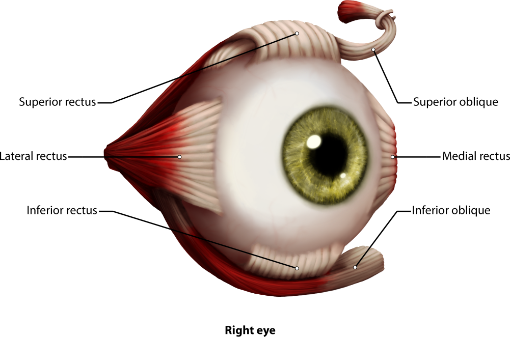
Figure 2: The eye and the six different extrinsic eye muscles. All muscles are colored reddish brown in the picture with white tendinous attachments to the eye. The superior rectus, lateral rectus, inferior rectus, inferior oblique, medial rectus and the superior oblique muscle as well as the trochlea are shown.
The muscles of the eye are innervated by three cranial nerves. Most of the muscles of the eye are innervated by the oculomotor nerve (cranial nerve III). The lateral rectus is innervated by the abducens nerve, CN IV, which causes abduction of the eye. The trochlear nerve, CN VI, innervates the superior oblique.
A Condition of the Eye Dealing with Eye Muscles
Strabismus (related to lazy eye) - Strabismus is a failure of the two eyes to maintain proper alignment and work together as a team. Some babies' eyes may appear to be misaligned, but are actually both aiming at the same object.
How to Test for Strabismus
Strabismus is classified by the direction the eye turns:
- Inward turning is called esotropia
- Outward turning is called exotropia
- Upward turning is called hypertropia
- Downward turning is called hypotropia.
You Use Cardinal Position of Gaze to Check the Tracking of These Positions
In ophthalmology the Cardinal Gaze tests one of six positions to which the normal eye may be turned. This test evaluates the functioning of the six extraocular muscles and the three corresponding cranial nerves. The positions and the corresponding muscles and nerves are:
- Straight Nasal: medial rectus and cranial nerve III
- Up Nasal: inferior oblique and cranial nerve III
- Down nasal: superior oblique and CN IV
- Straight temporal: lateral rectus and CN VI
- Up temporal: superior rectus and CN III
- Down Temporal: inferior rectus and CN III, also called extraocular movement.