Types of Muscle Tissues, Cellular Anatomy and the Functional Unit of Contraction (the Sarcomere)
Objectives
- Students will be able to compare the structural and functional characteristics and special properties of the three types of muscular tissue – Skeletal, Cardiac and Smooth.
- Students will be able to describe the relationship between the sarcolemma membrane and cellular ions.
If I were to ask you to point to a muscle, right now, most of you would point to one of your skeletal muscles like the biceps brachii, rectus abdominis, or even the gastrocnemius. While skeletal muscles are the most familiar, they are only one of the types of muscle that are found in your body. There are in fact 3 different types of muscles that are all structurally different from one another. Because of this difference in structure there is a distinct diversity in function. Simply put, the 3 different types of muscle all look different, so they have very different jobs.
Three Types of Muscle Structure
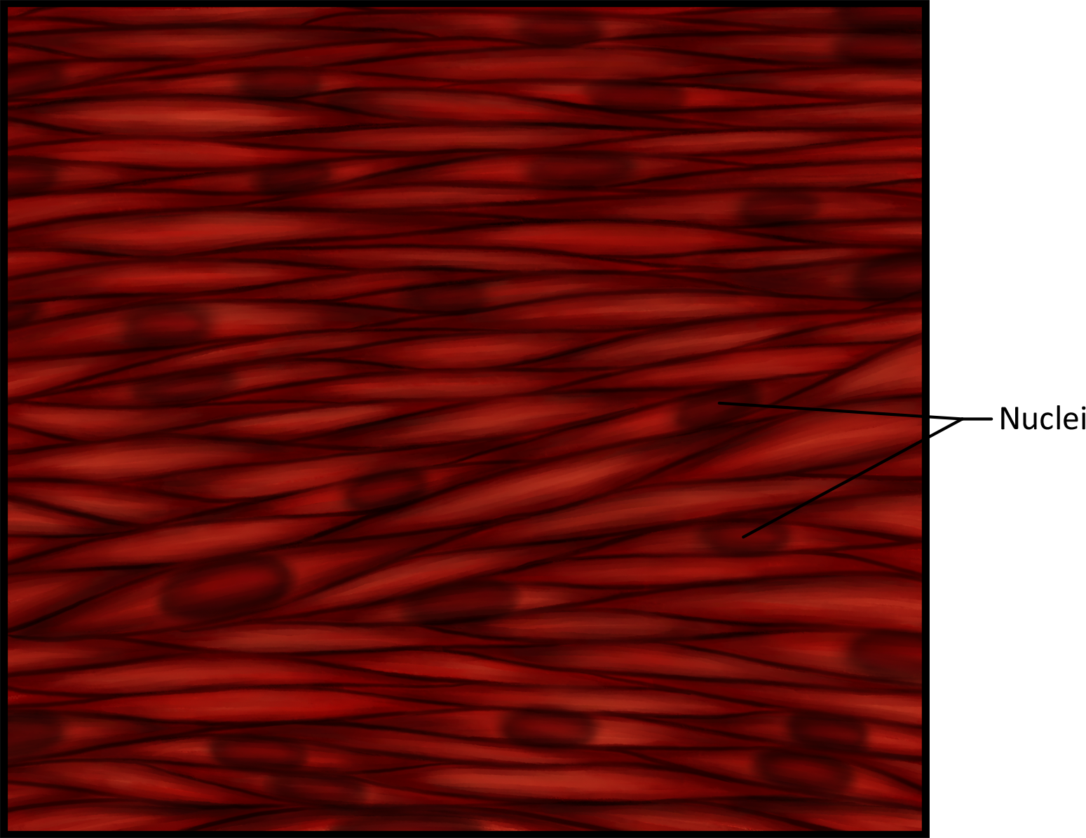
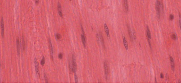
First, we have already mentioned skeletal muscle. Skeletal muscle is named because it moves your skeleton and we utilize this muscle mainly for locomotion and support of the body. This type of muscle is under voluntary control, which means that your brain controls the contraction. For instance, if you want to flex your arm and show off your biceps brachii, you can do so simply with a thought or intention of the movement; similar to: you think, therefore you do. These muscles are striated, or striped, due to the organization of intracellular proteins into units. Because the proteins are so highly organized and the units repeat, a striped appearance of this tissue results. These muscle cells are also multinucleated meaning that each cell has more than one nucleus. The biggest question asked here is WHY? Muscle cells are also known as muscle fibers, because they are extremely long cylindrical cells. One cell can run the entire length of a skeletal muscle, growing up to 30 cm in length (think of how long a giraffe's neck muscle fibers may be). With a cell being that expansive, there must be more than one control center (nucleus) to make sure the cell is functioning properly and maintaining homeostasis. During development, mesoderm differentiates into myoblasts (progenitor cells, each with its own nucleus) that then fuse together to create a larger, more elongated multinucleated skeletal muscle cell.
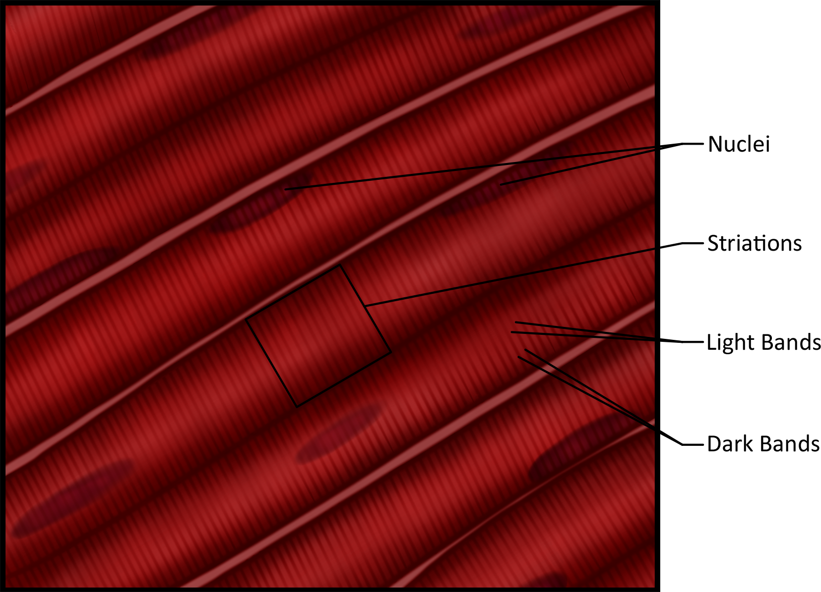
(a)
(b)
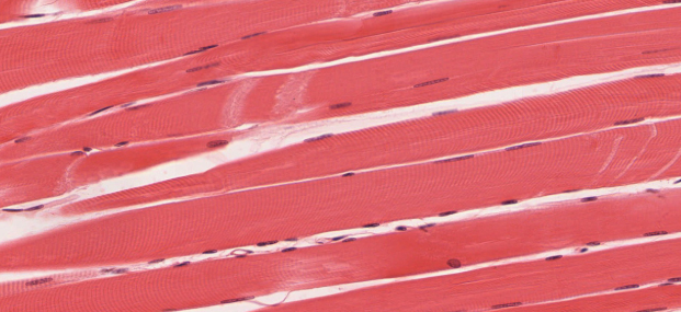
Figure 1. (a) This image depicts skeletal muscle cells; note the dark and light bands, striations and nuclei (b) An actual micrograph of skeletal muscle.
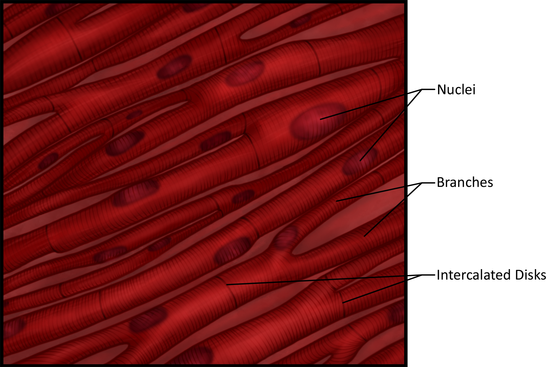
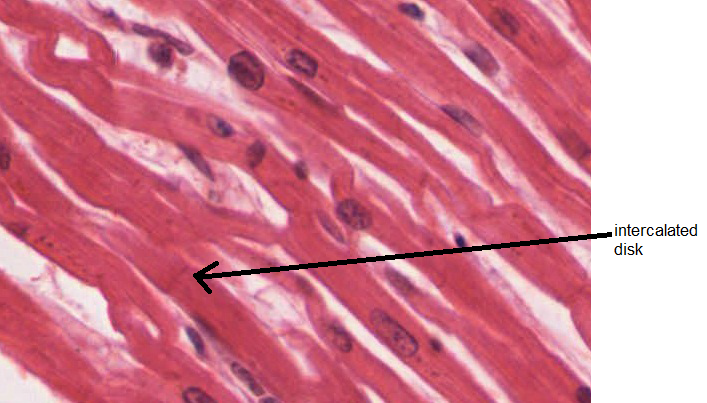
Cardiac muscle is named because it is found in the heart and we utilize this muscle to pump blood all over the body through the circulatory system. These muscle cells are also striated, like skeletal muscle, meaning that the organization of the proteins within a cardiac muscle cell mimics that found in a skeletal muscle cell. Due to these structural similarities, when you learn how a skeletal muscle contracts to cause movement you will then be able to adopt that knowledge to understanding cardiac muscle cell contraction. Even though there are similarities that exist between cardiac and skeletal muscle structure there are also great differences. Cardiac muscle cells are much shorter than skeletal muscle fibers, and because of this only have one nucleus instead of multiple. These cells are often branching creating a crisscross cell arrangement in the myocardium (cardiac muscle wall). This crisscross arrangement helps to effectively move the blood out of the heart into the awaiting blood vessels. These smaller cells must be connected so that they receive the proper signals and contract in a synchronized manner. The areas where 2 cells are joined are called intercalated discs and can be seen clearly in histology slide below. These discs contain desmosomes, junctions that hold the cells together (much like a zipper in a dress) and gap junctions, that allow the cells to directly communicate with each other.
Cardiac muscle is under involuntary control, meaning that you cannot control your heart rate to any great degree simply by thinking about it. The heart beats not because of conscious thought or intention but rather due to its own unique intrinsic properties and the autonomic nervous system. This is very beneficiamicrograph next to sketchl because as you sit reading this your heart continues to beat. If you had to tell it to beat each and every time it did so, you would not be able to enjoy this text or sleep at night without fear of missing several beats and incurring disaster. You will delve more into this fascinating phenomenon during the circulatory system chapter.
(a)
(b)
Figure 2. (a) This image depicts cardiac muscle cells; note the striations, branching nature and interacalated discs (b) An actual micrograph of cardiac muscle.
Lastly, smooth muscle fibers are named for their homogenous appearance, meaning that the cells look uniform throughout and do not have striations. This leads us to infer that because they are not organized in the same way that skeletal and cardiac muscle is organized, their method of contraction is different from what you will learn in this unit. Instead of the muscle cell shortening when it is stimulated, smooth muscle cells twist and bunch when stimulated. Visualize holding bread dough in your hands, placing one hand at either end of the dough. If you move your hands toward each other the dough shortens into rounded configuration - this is how skeletal and cardiac muscle contract. If you begin to twist the dough and move your hand toward each other forming a compacted spiral you have now mimicked how smooth muscle contracts. This is because the proteins responsible for contraction are not organized into repeating units but rather are located around the periphery of the cell like a net. The regulatory protein in smooth muscle is also different, utilizing calmodulin and myosin light-chain kinase instead of troponin.
Smooth muscle is widely distributed throughout the body and found primarily in the walls of hollow organs such as blood vessels, the digestive system, the iris of the eye, urinary bladder, etc. This muscle, like cardiac, is also under involuntary control and the function varies depending on the location of the smooth muscle. Remember, involuntary means that you cannot control the muscle actions to any great degree. If you stand in front of the mirror with an unchanging light source regardless of how hard you think, "pupil constrict," they will not do so.
(a)
(b)
Figure 3. (a) This image depicts smooth muscle cells; note the nuclei (b) An actual micrograph of smooth muscle.