Eyeball Structure
The eye is composed of three main layers or coats of tissues called tunics. The outermost layer is the fibrous tunic, which includes the white sclera and clear cornea. The sclera accounts for five sixths of the surface of the eye, most of which is not visible, though humans are unique compared with many other species in having so much of the "white of the eye" visible (Figure 3). The transparent cornea covers the anterior tip of the eye and allows light to enter the eye. This cornea is one of two major structures, along with the lens, in the eye that will cause light to bend and be focused on the posterior or back of the eye, where special types of receptors called photoreceptors are located. The middle layer of the eye is the vascular tunic, which is mostly composed of the choroid, ciliary body, and iris. The choroid is a layer of highly vascularized connective tissue that provides a blood supply to the eyeball. The choroid is posterior to the ciliary body, a muscular structure that is attached to the lens by zonule fibers (also called suspensory ligaments). These two structures bend the lens, allowing it to focus light on the back of the eye. Overlaying the ciliary body, and visible in the anterior eye, is the iris—the colored part of the eye. The iris is a smooth muscle that opens or closes the pupil, which is the hole at the center of the eye that allows light to enter. The iris constricts the pupil in response to bright light and dilates the pupil in response to dim light. The innermost layer of the eye is the neural tunic, or retina, which contains the nervous tissue responsible for photoreception.
The eye is also divided into two cavities: the anterior cavity and the posterior cavity. The anterior cavity is the space between the cornea and lens, including the iris and ciliary body. It is filled with a watery fluid called the aqueous humor. The anterior cavity is also divided up into two chambers: the anterior chamber and the posterior chamber. The posterior chamber contains the lens and the ciliary bodies. It is at the ciliary bodies that the secretion of the aqueous humor take places derived from capillaries in their walls. The aqueous humor then circulates into the anterior chamber through the pupil and is then returned to the blood through the scleral venous sinus, that is a small opening found at the junction of the cornea and sclera. This opening allows for the drainage of aqueous humor from the anterior compartment of the eye and, therefore, reducing intraocular pressure related to glaucoma.The posterior cavity is the space behind the lens that extends to the posterior side of the interior eyeball, where the retina is located. The posterior cavity (vitreous chamber) is filled with a more viscous fluid called the vitreous humor.
The retina is composed of several layers and contains specialized cells for the initial processing of visual stimuli. The photoreceptors (rods and cones) change their membrane potential when stimulated by light energy. The change in membrane potential alters the amount of neurotransmitter that the photoreceptor cells release onto bipolar cells in the outer synaptic layer. It is the bipolar cell in the retina that connects a photoreceptor to a retinal ganglion cell (RGC) in the inner synaptic layer. There, amacrine cells additionally contribute to retinal processing before an action potential is produced by the RGC. The axons of RGCs, which lie at the innermost layer of the retina, collect at theoptic disc and leave the eye as the optic nerve (see Figure 3). Because these axons pass through the retina, there are no photoreceptors at the very back of the eye, where the optic nerve begins. This creates a "blind spot" in the retina, and a corresponding blind spot in our visual field.
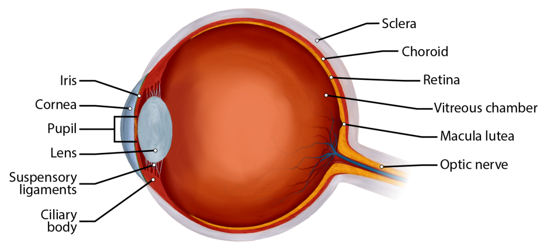
Figure 3: This picture shows a section through an eye showing most of the major parts of the eye such as the sclera, cornea, choroid, retina, the macula lutea (with the fovea in the center), optic nerve, suspensory ligaments, lens, iris, pupil, ciliary bodies, and the two major cavities of the eye (anterior cavity, anterior to the lens, and vitreous chamber or posterior cavity).
Now we will examine the microscopic level of the retina.