

Autonomic Reflexes and Hypothalamic Control
Objectives:
By the end of this section the student will be able to:
- Describe the components of an autonomic reflex and the importance of the hypothalamus to the ANS.
Can you answer these questions?
- What are the components of an autonomic reflex compared to a somatic reflex?
- How does the hypothalamus control the ANS?
The autonomic nervous system regulates organ systems through circuits that resemble the reflexes described in the somatic nervous system. Earlier in this module we discussed the similarities and difference that exist between the SNS and the ANS. The key difference between the divisions was conscious control – is the effector voluntarily controlled by thought or intention. This difference is useful when trying to decipher if a reflex is somatic or autonomic in nature. The example that is most used and confused is breathing. The question I pose to you is, "Is breathing a somatic or autonomic reflex?"
The correct answer is somatic reflex. Most students are confused because breathing occurs most often without your thinking about it. BUT just because you don't think about breathing every 5 seconds and it occurs anyway does not make it an autonomic response. If you wanted to take a deep breath in could you do so with thought and intention? Try it! The diaphragm is a skeletal muscle that be controlled with thought/intention therefore demonstrating that the effector of the reflex is controlled voluntarily. This is why breathing is a somatic reflex.
There are other differences that exist, such as structural differences in the reflex arc and the effectors of each division. Somatic responses are solely based on skeletal muscle contraction. The autonomic system, however, targets cardiac and smooth muscle, as well as glandular tissue.
One structural difference between a somatic reflex and an autonomic reflex (or visceral reflex) is in the efferent branch and is one that we are already familiar with. The motor output of a somatic reflex is the lower motor neuron that begins in the anterior horn of the spinal cord and projects directly to a skeletal muscle to cause its contraction. The output of an autonomic reflex is a two-step pathway starting with the preganglionic fiber emerging from a lateral horn neuron in the spinal cord, or the brain stem, to a ganglion—followed by the postganglionic fiber projecting to a target effector. The other part of a reflex, the afferent branch, is often the same between the two systems. Sensory neurons receiving input from the periphery—with cell bodies in the sensory ganglia, either of a cranial nerve or a dorsal root ganglion adjacent to the spinal cord—project into the CNS to initiate the reflex (Figure 12).
Helpful Study Fact 3: The Latin root "effere" means "to carry." Adding the prefix "ef-" suggests the meaning "to carry away," whereas adding the prefix "af-" suggests "to carry toward or inward."
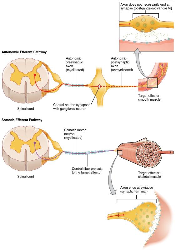
Figure 12: The afferent inputs to somatic and visceral reflexes are essentially the same, whereas the efferent branches are different. Somatic reflexes, for instance, involve a direct connection from the ventral horn of the spinal cord to the skeletal muscle. Visceral reflexes involve a projection from the central neuron to a ganglion, followed by a second projection from the ganglion to the target effector.
Another difference that exists between a somatic and autonomic reflex is involvement of the CNS. Somatic reflexes involve sensory neurons that connect sensory receptors to the CNS and motor neurons that project back out to the skeletal muscles. Autonomic reflexes that involve the thoracolumbar or craniosacral systems share similar connections. However, there are reflexes that do not need to involve any CNS components. A long reflex has afferent branches that enter the spinal cord or brain and involve the efferent branches, as previously explained. A short reflex is completely peripheral and only involves the local integration of sensory input with motor output in a peripheral ganglion (Figure 13).
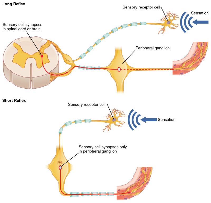
Figure 13: Sensory input can stimulate either a short or a long reflex. A sensory neuron can project to the CNS or to an autonomic ganglion. The short reflex involves the direct stimulation of a postganglionic fiber by the sensory neuron, whereas the long reflex involves integration in the spinal cord or brain.
The difference between short and long reflexes is in the involvement of the CNS. Somatic reflexes always involve the CNS, even in a monosynaptic reflex in which the sensory neuron directly activates the motor neuron (for example, the knee reflex, which only goes through the spinal cord since it only needs one synapse). That synapse is in the spinal cord or brain stem, so it has to involve the CNS. However, in the autonomic system there is the possibility that the CNS is not involved. Because the efferent branch of a visceral reflex involves two neurons—the central neuron and the ganglionic neuron—a "short circuit" can be possible. If a sensory neuron projects directly to the ganglionic neuron and causes it to activate the effector target, then the CNS is not involved.
The hypothalamus is the source of most of the central control of autonomic function. It regulates both autonomic function and endocrine function. It receives input from cerebral structures and projects to brain stem and spinal cord structures to regulate the balance of sympathetic and parasympathetic input to the organ systems of the body. The main pathways for this are the medial forebrain bundle and the dorsal longitudinal fasciculus.These two tracts connect the hypothalamus with the major parasympathetic nuclei in the brain stem and the preganglionic (central) neurons of the thoracolumbar spinal cord. The hypothalamus also receives input from other areas of the forebrain through the medial forebrain bundle. The olfactory cortex, the septal nuclei of the basal forebrain, and the amygdala project into the hypothalamus through the medial forebrain bundle. These forebrain structures inform the hypothalamus about the state of the nervous system and can influence the regulatory processes of homeostasis. A good example of this is found in the amygdala, which is found beneath the cerebral cortex of the temporal lobe and plays a role in our ability to remember and feel emotions.
