Skeletal Muscle Organization
Now that we have examined all the major tissues found in this complex organ we must now learn about how the organ is organized. Like the rest of the structures of your body, skeletal muscles are not simply thrown together, they are highly structured. Why is this organization so important? Two reasons, the organization of the intracellular proteins allows for the greatest contraction possible and the compartmentalization allows for activation of only the skeletal muscle cells needed for a particular force production. You wouldn't use the same force to lift a coffee mug as you would a couch. The compartments allow us to activate only a few muscle cells to lift the cup and then activate many more to lift the couch.
When we start to examine the structure of a whole skeletal muscle it is important to deconstruct the organ one layer at a time and after it is fully deconstructed, piece it back together from the smallest pieces into the whole organ. If you were to look at a whole skeletal muscle, like the biceps brachii, you would see that it is covered by a layer of connective tissue called the epimysium. This epimysium not only covers the entire skeletal muscle keeping its integrity as it contracts and extends, but also separates the skeletal muscle from other surrounding skeletal muscles allowing it to move independently. The epimysium also extends out and connects with the periosteum of the skeletal system to form tendons. This tendon ensures that when the skeletal muscle contracts it will pull on the attached bone and cause movement.
Next we cut the skeletal muscle along a transverse plane to see the inside of the organ. What we notice are many large bundles inside the skeletal muscle. These large bundles are called fascicles and are covered with a layer of connective tissue called the perimysium. The perimysium allows the nervous system to selectively activate muscle cells based on the need for a particular amount of force – remember the coffee mug and couch example?
If we look closely at each fascicle we will see many bundles inside of this structure that we call skeletal muscle cells or muscle fibers. Remember, both terms are widely used and acceptable. On the outside of the actual muscle cell sits that last layer of connective tissue called the endomysium. The endomysium surrounds the skeletal muscle cell separating it from other muscle cells nestled in the same fascicle. Underneath the endomysium is the cell membrane of this muscle cell/fiber itself, we call it the sarcolemma (means: muscle covering).
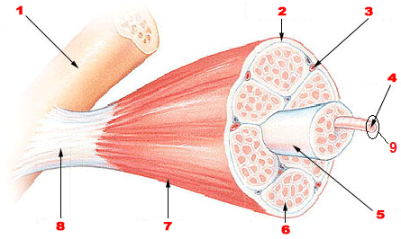
Figure 1. Skeletal Muscle Organization. Image thoroughly described below.
Figure 1 Long Description
- Bone with periosteum (structure in upper left hand corner, showing muscle attachment)
- Perimysium (connective tissue surrounding fascicles)
- Blood vessel (red, transports blood through muscle)
- Muscle fiber (long, cylindrical, multinucleate cells shown here sticking out of fascicle)
- Fascicle (bundle of muscle fibers within a skeletal muscle)
- Endomysium (connective tissue surrounding fibers)
- Epimysium (connective tissue around whole skeletal muscle)
- Tendon (extension from the periosteum, anchors muscle to the bone)
- Sarcolemma (membrane surrounding muscle fibers)
Within the sarcolemma are all of the cellular components in the sarcoplasm, but what stands out about these cells are that the majority of the cell is made of more bundles. The nuclei are pushed to the outside of the cell while the mitochondria are squeezed into available space between the bundles. These bundles are called myofibrils and are collections of proteins highly organized into groups called sarcomeres. Sarcomeres are the functional unit of skeletal muscle – meaning they are the smallest unit that can perform the functions of a skeletal muscle. Remember that these repeating sarcomeres give the skeletal muscle a striated appearance. We will discuss this more in a later section.
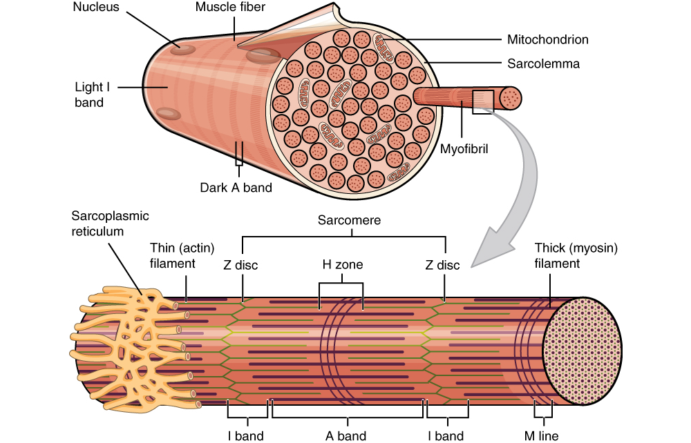
Figure 2. At this point in the module, you are encouraged to pay attention to the right portion of the image depicting a a myofibril protruding from a sarcolemma-surrounded muscle fiber. The myofibril is composed of repeating sarcomere units, depicted in an enlarged form below the grey arrow. Sarcomere compostition will be described in greater detail further in to this module.