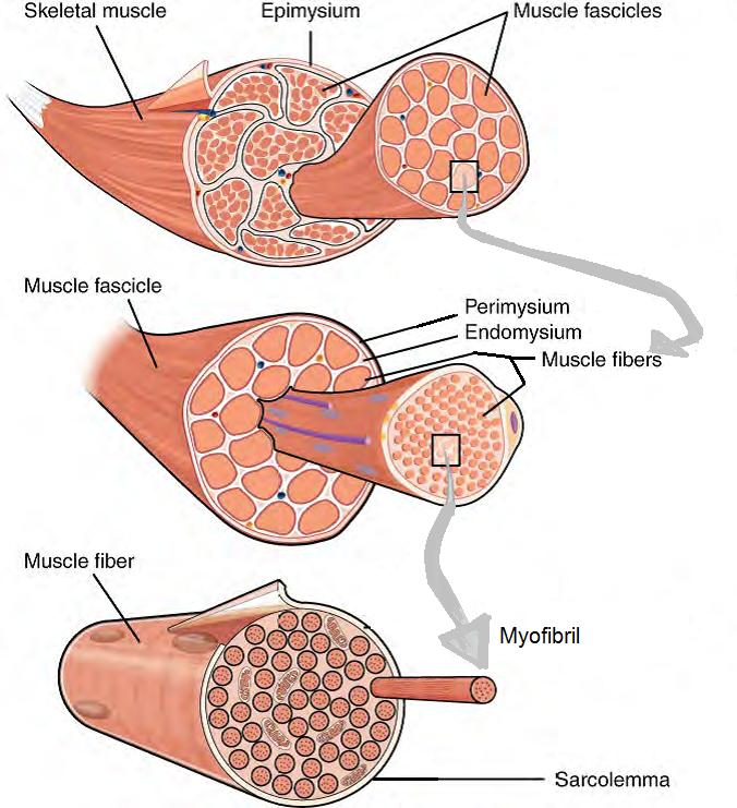
Figure 3. The Three Connective Tissue Layers. The entire skeletal muscle is covered by epimysium. Bundles of muscle fibers, called fascicles, are covered by the perimysium. Individual muscle fibers are covered by the endomysium.
Now that we have deconstructed the muscle, let's reconstruct it! Proteins are organized into repeating sarcomeres and then bundled into myofibrils. Myofibrils are bundled into skeletal muscle cells that are contained by a cell membrane known as the sarcolemma. On top of the skeletal muscle cell is the endomysium. Many of these skeletal muscle cells covered by their individual endomysium are bundled into a fascicle. The fascicle is covered with perimysium. Many fascicles are bundled together to create a whole skeletal muscle. This skeletal muscle is then covered with epimysium. If you are a visual learner seeing this organization in a flow chart may be helpful.
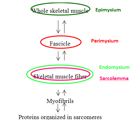
Figure 4. Muscle Fiber Organization Chart. This flow chart depicts the organization of a whole skeletal muscle as it is dissected from whole to its components. The coverings of each layer are denoted by colored circles. The starting point is a whole skeletal muscle covered by a green circle representing as the epimysium. If you were to cut a whole skeletal muscle you would see large groups known as fascicles covered by red circle representing as the perimysium. Looking at each fascicle you will find more groups known as skeletal muscle cells covered by a outer green and an inner pink circle, representing the endomysium and the sarcolemma respectively. Each skeletal muscle cell contains groups known as myofibrils which are composed of sarcomeres. You can reassemble the whole skeletal muscle from sarcomeres by reversing the arrow directionality.
![]()
Video 1. View the YouTube video Muscle Fiber Organization (opens in new window)
Looking more closely at the skeletal muscle fiber we can see defined repeating units that we know as sarcomeres. The sarcomere is defined by a Z disc on either end; the Z disc is a lattice work of protein filaments that mark the boundary between adjacent sarcomeres. Simply stated, a sarcomere extends from Z disc to Z disc. The center of the sarcomere is designated by the M line (M = middle).
The sarcomere itself is composed of 6 different protein molecules that can be classified into 3 groups based on its job in muscle contraction.
- Contractile proteins → actin and myosin
- Structural proteins → nebulin and titin
- Regulatory proteins → troponin and tropomyosin
The contractile proteins are the actual molecules that participate in muscle contraction. One of the contractile proteins is called actin or more commonly called the thin filament. Actin filaments are attached to the Z disc at each end of the sarcomere and radiate inward toward the M line. The actin does not connect in the middle it simply points toward the other actin filament. If you look closely at the actin filament you will see that the filament is actually made out of many much smaller spherical actin subunits. Each actin subunit has a binding site on it for the other contractile protein, myosin. The actin subunits are placed in a double helical pattern around a center structural protein, nebulin. We call actin the thin filament due to its small width only composed of 2 strands of actin subunits and 1 nebulin protein.
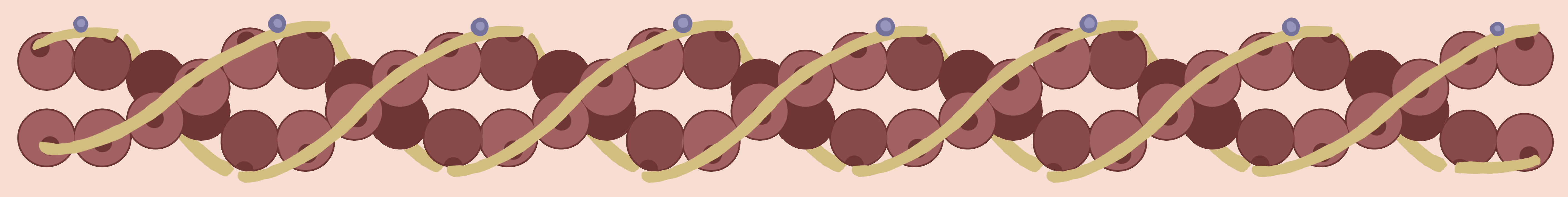
Figure 5. Actin myofilaments contain globular proteins called G-actin molecules (brown balls) that are surrounded by strands of tropomyosin molecules (yellow strands). The troponin protein molecules (blue circles) regulate the movement of the tropomyosin in the presence of calcium.
The second contractile protein is myosin, or more commonly, the thick filament. Myosin filaments are centered at the M line of the sarcomere and extend toward either Z disc without fully reaching them. Titin is the structural protein associated with myosin. The titin is a coiled structural protein that inserts at the Z discs and extends through the middle of the sarcomere. The myosin is anchored to the titin so that it stays unwavering in the middle of the sarcomere when contraction happens instead of shifting in either direction toward a Z disc. If this was to happen damage to the sarcomere would result. If you look closely at the myosin filament you will see it is very thick because the filament is made of many myosin proteins that are wound together. Each individual myosin protein resembles a golf club and has a head portion, a neck/hinge portion, and a tail portion. It is the tail portion of the myosin protein that intertwines with other myosin proteins to create the filament. The head of the myosin protein sticks out of the bundle and can pivot in multiple directions. This head portion is also the part that binds to the binding site on the actin subunit mentioned above.
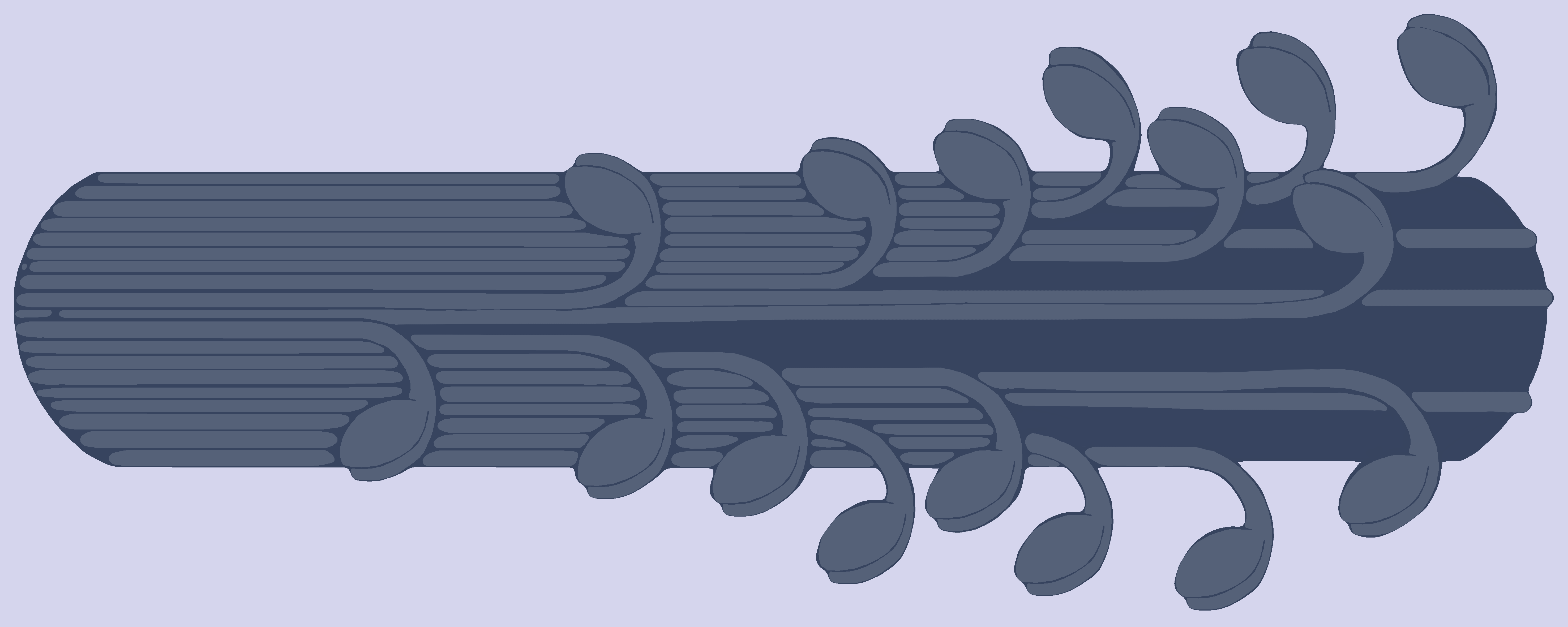
Figure 6. The Myosin filament. Each individual myosin protein resembles a golf club and has a head portion, a neck/hinge portion, and a tail portion. It is the tail portion of the myosin protein that intertwines with other myosin proteins to create the filament (the bundle of golf clubs). The head portion is the part that binds to the myosin binding site on the G-actin protein.
When actin and myosin are arranged in the sarcomere, as seen below, there are various bands and zones that are created. These are labeled and important to know because each band or zone contains a different framework of contractile proteins and therefore refracts light differently. The H zone is located around the M line and extends from one end of the actin filament to the opposing end of the actin filament. This means that the H zone contains only myosin filaments. On an electron micrograph this area appears light grey with a dark line down the center (M line). The I band is located around the Z disc and extends from one end of the myosin filament of one sarcomere to the opposing end of the myosin filament in the NEIGHBORING sarcomere. This means that the I band contains actin filaments only. On an electron micrograph this area appears almost white with a dark line down the center (Z disc). The area is lighter than the H zone because actin is thinner than myosin. The last band is the A band and it extends from one end of the myosin filament of one sarcomere to the opposing end of the myosin filament in the SAME sarcomere. This band is the area of overlap (A = Area of overlap) and contains both actin and myosin filaments. On an electron micrograph this area appears dark grey due to the presence of both filaments with the H zone in the middle of the A band. [Think of the A-band as the d A rk band and the I-band as the l I ght band.] So a sarcomere shows the coloration from one Z disc to the other: white, dark grey, light grey, dark grey, white. If you take your pencil and shade in this pattern what do you see?
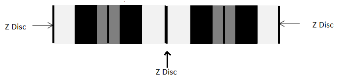
Figure 7. Sarcomere with Z Discs already labeled. In the text below, you will learn about the rest of the bands and zones.
Stripes!!! Remember we call these striations in muscle cells. Also, remember that sarcomeres repeat and if you continue the pattern above the striations become very evident. When the muscle cell is stimulated to contract, the actin is pulled toward the M line by the myosin filaments. It is important to remember that the contractile filaments DO NOT SHORTEN they simply slide past each other toward the middle which shortens the sarcomere, therefore shortening the muscle cell, and fascicle, and whole skeletal muscle. This mode of filament movement is explained later in the Sliding Filament Theory - note how the name describes what takes place.
The question to you is: Which of the bands and zones that we just discussed shorten during contraction?
- A band?
- H zone?
- I band?
- Multiple ones listed?
The correct answer is the H zone and I band. Remember, the A band extends from one end of the myosin filament to the other end of the filament in the SAME sarcomere, and since these thick filaments don't shorten during contraction - the length of this band won't change. However, the H zone consists of myosin only and the I-band of actin only. During contraction the filaments begin to slide and overlap more which means the areas where only one type of filament exists shortens substantially. The way I have always remembered this was that "HI" is a shorter version of "Hello" therefore during contraction the H and I shorten.
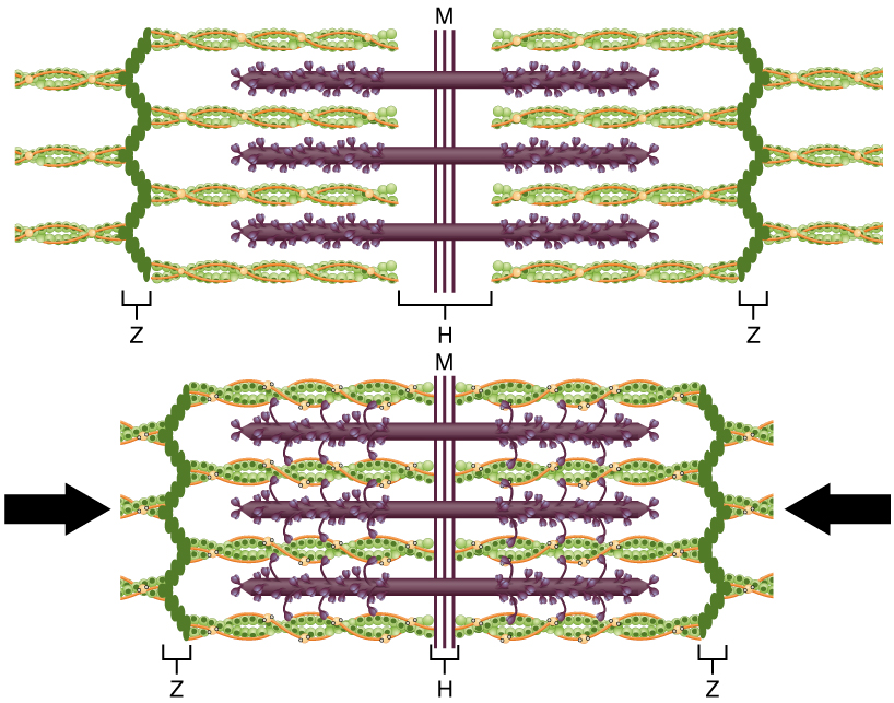
Figure 8. The Sliding Filament Model of Muscle Contraction. When a sarcomere contracts, the Z lines (depicted here by vertical pleated chains of dark green circles) move closer together, and the I band becomes smaller. The A band (shown here as the most contracted width of the H-zone) stays the same. At full contraction, the thin (shown here as twisted green and orange strands) and thick filaments (shown here as purple strands with protrustions) overlap.
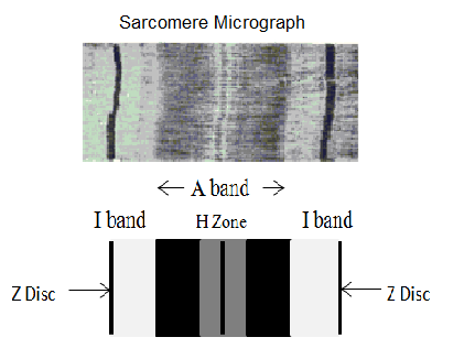
Figure 9. The top image is of an actuall saromere micrograph, while the bottom image provides helpful labels of the (outermost, darkest) Z Disc; (lighter, inside) I band and the (central) A band which encompasses the (lighter) H Zone in the very center. More detailed written descriptions of all of these terms can be found in the glossary.
![]()
Video 2. View the YouTube video Sarcomere Analogy (opens in new window)
Skeletal Muscle Structure
Activity not available on mobile devices (description)
Sarcomere
Activity not available on mobile devices (description)