

Vision
Objectives
By the end of this module you will be able to:
- List the supporting structures around the eye and a disorder called Stabismus
- Describe the structure of the eyeball
- Describe the structures responsible for the sense of vision
- Describe the processes of phototransduction
Vision is a special sense that involves the transduction (conversion) of wavelengths or particles of light called photons into nerve impulses at the eye, sending these impulses to the brain and then interpreting these impulse into images by the visual cortex of the brain. The eyes are located in the orbital sockets of the skull and surrounded by a series of accessory structures that include the eyelids, eyelashes, muscles, lacrimal (tear) glands, and mucous membranes. The eyelids, with the eyelashes, help to protect the eye from damage by blocking material from entering the eye. The inner part of the eyelid as well as the outer part of the eye is lined with a membrane called the conjunctiva. The part of the conjunctiva that lines the eyelid is called the palpebral conjunctiva. This then connects the eyelids to the eyeball. On the lateral edge of the eye is the lacrimal gland that produces tears. Tears flow through the lacrimal duct to the inner corner of the eye washing foreign particles from the eye, where they drain into a duct that connects to the nasal cavity. In addition to cleansing the eye, these tears also contain an enzyme called lysozyme that helps to protect the eye against infections.
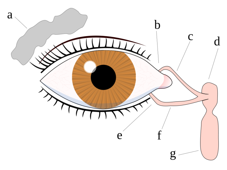
Figure 1: The Lacrimal apparatus and the drainage of tears start with the lacrimal gland in the upper side of the eye to the lower corner of the eye through which tears drains into the nasal cavity. The lacrimal gland in the upper side of the eye is colored gray while the lacrimal apparatus consisting of the canals and the nasolacrimal duct are colored pink. a. tear gland / lacrimal gland, b. superior lacrimal punctum/opening, c. superior lacrimal canal, d. tear sac / lacrimal sac, e. inferior lacrimal punctum, f. inferior lacrimal canal, g. nasolacrimal canal