Parasympathetic Neurons
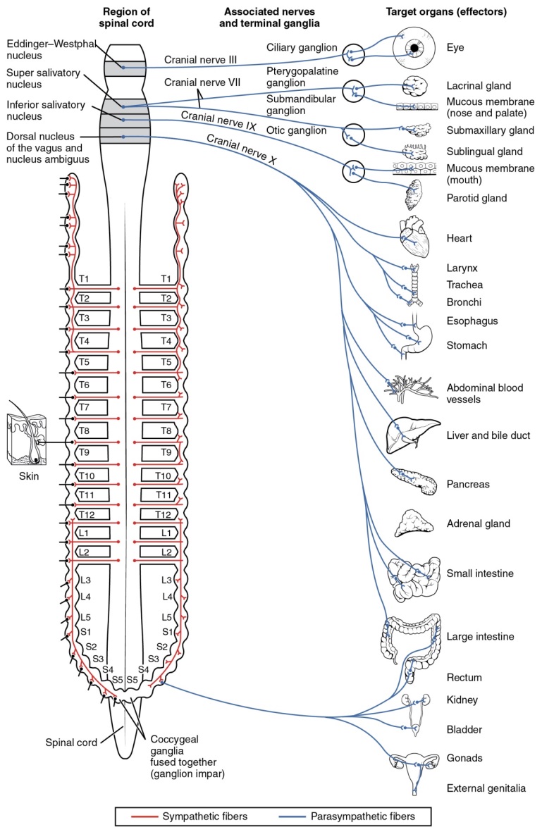
A diagram that shows the connections of the parasympathetic system is somewhat like a circuit diagram that shows the electrical connections between different receptacles and devices. In Figure 7, the "circuits" of the parasympathetic system are intentionally simplified.

Figure 7: Neurons from brain-stem nuclei, or from the lateral horn of the sacral region of the spinal cord, project to terminal ganglia near or within the various organs of the body. Axons from these postganglionic neurons then project the short distance to those target effectors. Notice the length of the pre and postganglionic neurons because of the location of the autonomic ganglion to the spinal cord.
As we have previously stated: an axon from the neuron originating from the CNS that projects to a autonomic ganglion is referred to as a preganglionic fiber or neuron, and represents the output from the CNS to the ganglion. Because the parasympathetic ganglia are terminal or located near the target effector, preganglionic sympathetic fibers are relatively long. The preganglionic fiber passes the signal across the synapse in the autonomic ganglion by releasing the ACh. The neurotransmitter diffuses across the synaptic cleft and binds to nicotinic ACh receptors on the postganglionic fiber, thus stimulating the neuron. The postganglionic fiber or neuron —the axon from a ganglionic neuron that projects to the target effector—represents the output of a ganglion that directly influences the organ. Compared with the preganglionic fiber, postganglionic sympathetic fibers are short because of the short distance from the ganglion to the target effector. The postganglionic fiber passes the signal across the synapse to the effectors by releasing the ACh. The neurotransmitter diffuses across the synaptic cleft and binds to muscarinic ACh receptors on the effector, thus causing systemic change. Muscarinic receptors are similar to nicotinic receptors because they bind to ACh but unlike nicotinic receptors they operate via a G protein cascade instead of an ion channel. This means that instead of ACh simply opening channels and activating the postganglionic fiber, a chain of events is set in, motion by the binding of ACh and those events eventually activate the postganglionic fiber. Because ACh is the neurotransmitter used at the effector, the overall response of this system is termed cholinergic. (acetylCHOLINE = CHOLINErgic).

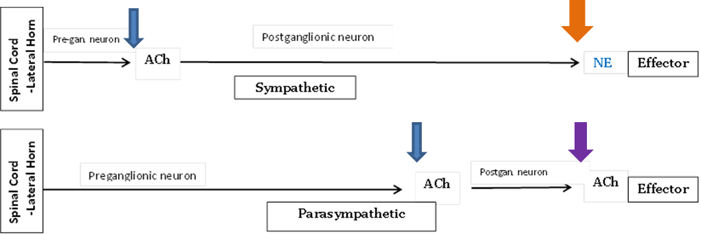
Figure 8: In this figure of the ANS pathways, thinner blue arrows show where nicotinic receptors receive acetylcholine (ACh) at the postganglionic neuron, the orange arrow shows where adrenergic alpha and beta receptors receive Norepinephrine (NE) along the sympathetic pathway. The purple arrow shows where muscarinic receptors receive the ACh at the effector.
Return to Table 2 to review these Neurotranmitters and receptors for the Parasympathetic Division.
So you can understand that this "rest and digest" division of the ANS would have what is often called cholinergic responses such as: increase digestive functions, increase pancreatic secretions of digestive enzymes, relax sphincters, increase defecation and urination, too, along with other less stressful functions.
SLUDD is as an acronym used to describe the heightened responses of the parasympathetic nervous system:
- Salivation (increased)
- Lacrimation (increased)
- Urination (increased)
- Digestion (increased)
- Defecation (increased)
… and 3 decreases (in the rate and force of the heartbeat, airway size and rate of breathing, and pupil size)
… and in the sex organs vasodilation for erection of the female clitoris and the male penis.
You can understand why this division is also called "feed and breed" as well as "rest and digest" as nicknames.