

Venous Return
After passing through the CNS, blood returns to the circulation through a series of dural sinuses and veins mentioned before. The superior sagittal sinus runs in the groove of the longitudinal fissure, where it absorbs CSF from the meninges.

Figure 16. Dural Sinuses and Veins Blood drains from the brain through a series of sinuses that connect to the jugular veins. You will need to learn these for the Blood vessels module later.
Ventricles (sacs of fluid)
Another aspect of the adult CNS structures that relates to embryonic development is the ventricles—open spaces within the CNS where cerebrospinal fluid is produced and circulates. They are the remnant of the hollow center of the neural tube. Each part of the adult brain is associated with one of the four ventricles that contain cerebrospinal fluid (CSF). Two ventricles, the largest of the four, are wing-like and have protruding posterior horns. These two lateral ventricles (right and left) sit on opposite sides of the septum pellucidum in each cerebral hemisphere. A narrow third ventricle sits between the two thalamic halves in the diencephalon. A hole, or interventricular foramen, allows fluid to drain from each of the lateral ventricles into the single third ventricle. From the third ventricle the CSF goes through the cerebral aqueduct to a fourth ventricle. The fourth, inferior ventricle sits anterior to the cerebellum and posterior to the pons, which then drains into the central canal of the spinal cord and through openings (median and lateral apertures) into the subarachnoid space around the brain and spinal cord surfaces.
The CSF is formed from modified blood vessels found in each ventricle called choroid plexuses. These networks of blood vessels are lined with ependymal cells (ĕ-pen'-dy-mal) to filter blood plasma to form CSF, which will contain nutrients and oxygen for the neurons and neuroglial cells. Because the blood is continually filtered here, the CSF continuously circulates through the ventricles and subarachnoid space. Within the subarachnoid space, the CSF flows around all of the CNS, providing two important functions. As with elsewhere in its circulation, the CSF picks up metabolic wastes from the nervous tissue and moves it out of the CNS. It also acts as a liquid cushion for the brain and spinal cord. By surrounding the entire system in the subarachnoid space, it provides a thin buffer around the organs within the strong, protective dura mater. The arachnoid granulations are outpockets of the arachnoid membrane into the dural sinuses (large veins in the dura mater) so that CSF can be reabsorbed into the blood, along with the metabolic wastes. From the dural sinuses, blood drains out of the head and neck through the jugular veins, along with the rest of the circulation for blood, to be reoxygenated by the lungs and wastes to be filtered out by the kidneys. Similar to clinical blood work, a sample of CSF (via the lumbar puncture) can be withdrawn to find chemical evidence of neuropathology or metabolic traces of the biochemical functions of nervous tissue. You will follow the path of CSF through the brain and spinal cord in lab.
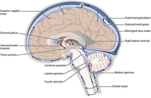
Figure 17.
View the Cerebrospinal Fluid Circulation video (opens in a new window) that shows the flow of CSF through the brain and spinal cord, and how it originates from the ventricles.

