The Myth of Left Brain/Right Brain
There is a persistent myth that people are "right-brained" or "left-brained," which is an oversimplification of an important concept about the cerebral hemispheres. There is some lateralization of function, in which the left side of the brain is devoted to language function and the right side is devoted to spatial and nonverbal reasoning. Whereas these functions are predominantly associated with those sides of the brain, there is no monopoly by either side on these functions. Many pervasive functions, such as language, are distributed globally around the cerebrum.
Some of the support for this misconception has come from studies of split brains. A drastic way to deal with a rare and devastating neurological condition (intractable epilepsy) is to separate the two hemispheres of the brain. After sectioning the corpus callosum, a split-brained patient will have trouble producing verbal responses on the basis of sensory information processed on the right side of the cerebrum, leading to the idea that the left side is responsible for language function.
However, there are well-documented cases of language functions lost from damage to the right side of the brain. The deficits seen in damage to the left side of the brain are classified as aphasia, a loss of speech function; damage on the right side can affect the use of language. Right-side damage can result in a loss of ability to understand figurative aspects of speech, such as jokes, irony, or metaphors. Nonverbal aspects of speech can be affected by damage to the right side, such as facial expression or body language, and right-side damage can lead to a "flat affect" in speech, or a loss of emotional expression in speech—sounding like a robot when talking.
The Diencephalon (di-en-SEF-a-lon)
The diencephalon is the connection between the cerebrum and the rest of the nervous system, with one exception. The rest of the brain, the spinal cord, and the PNS all send information to the cerebrum through the diencephalon. Output from the cerebrum passes through the diencephalon. The single exception is the system associated with olfaction, or the sense of smell, which connects directly with the cerebrum.
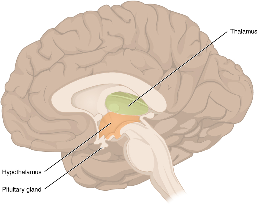
The diencephalon is deep beneath the cerebrum and makes up the walls of the third ventricle. The diencephalon can be described as any region of the brain with "thalamus" in its name. The two major regions of the diencephalon are the thalamus itself and the hypothalamus (see figure below). There are other structures, such as the epithalamus, which contains the pineal (`pin-ē-al) gland, or the subthalamus, which includes the subthalamic nucleus that is part of the basal nuclei.
Thalamus
The thalamus is a collection of nuclei that relay information between the cerebral cortex and the PNS, spinal cord, or brain stem. All sensory information, except for the sense of smell, passes through the thalamus before processing by the cortex. Axons from the peripheral sensory organs (first order neuron) synapse in the thalamus, and thalamic neurons (second order neuron) project directly to the cerebrum. The thalamus acts like a relay station or a receptionist asking "How may I direct your call?" and sending the incoming information from the first order neuron to the correct location on the cortex via the second order neuron. The thalamus does not just pass the information on, it also processes that information. For example, the portion of the thalamus that receives visual information will influence what visual stimuli are important, or what receives attention.
The cerebrum also sends information down to the thalamus, which usually communicates motor commands. This involves interactions with the cerebellum and other nuclei in the brain stem. The cerebrum interacts with the basal nuclei, which involves connections with the thalamus.
Hypothalamus
Inferior and slightly anterior to the thalamus is the hypothalamus, the other major region of the diencephalon. The hypothalamus is a collection of nuclei that are largely involved in regulating homeostasis. The hypothalamus is the executive region in charge of the autonomic nervous system and the endocrine system through its regulation of the anterior pituitary gland. Other parts of the hypothalamus are involved in memory and emotion as part of the limbic system.

Figure 9. The diencephalon is composed primarily of the thalamus and hypothalamus , which together define the walls of the third ventricle. The thalami are two elongated, ovoid structures ("bird's head") on either side of the midline that make contact in the middle. The hypothalamus is inferior and anterior to the thalamus, culminating in a beak-like sharp angle to which the pituitary gland is attached. Above the diencephalon, the corpus callosum and the fornix can be seen (similar to crests on a bird's head).
The thalami are two elongated, ovoid structures on either side of the midline that make contact in the middle - at the "eye" of the bird's head called the Intermediate Mass of the thalamus. The hypothalamus is inferior and anterior to the thalamus, culminating in a "beak-like" structure to which the pituitary gland is attached.
The limbic system is a connected set of structures between the forebrain and hindbrain that regulates emotion, as well as behaviors related to fear and motivation. It plays a role in memory formation and includes parts of the thalamus and hypothalamus as well as the hippocampus. One important structure within the limbic system is a temporal lobe structure called the amygdala, mentioned earlier . The two amygdala (one on each side) are important both for the sensation of fear and for recognizing fearful faces. As aforementioned the amygdala develops early in life and persists as the lead emotional decision-maker until the prefrontal cortex develops fully in the mid-20s.


