
![]()
![]()
Animation 2. The Brain Triggers an Action Potential. View the YouTube animation Muscle Excitation Step 1. (opens in new window)
![]()
![]()
Animation 3. Action Potential Arrives at the Muscle Cell. View the YouTube animation Muscle Excitation Step 2. (opens in new window)
Now that ACh has been released into the synaptic cleft, it will diffuse through the cleft to the surface of the skeletal muscle cell which is called the sarcolemma. The portion of the sarcolemma that faces the synaptic end bulb is folded many times to increase surface area and have more receptors for ACh. This folded portion is called the motor end plate. Remember from previous chapters that the folding increases surface area which in this case is crucial so that you can pack more ACh receptors into this area and will need less ACh to pass the signal along to the muscle that it is now time to contract. Let's draw this out quickly to make sure it is clear. Draw a straight line and a squiggly line up and down. Now add an ACh receptor every centimeter in your drawing. How many receptors did you fit on the straight line versus the folded line?
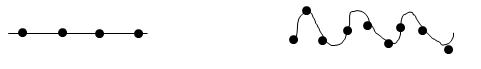
Figure 1. Folded lines have more surface area than straight lines.
The ACh diffuses through the synaptic cleft and binds to one of the many ACh receptors present on the motor end plate of the sarcolemma. These receptors are also known as ligand-gated sodium channels. This means when the chemical ligand is present (in this case ACh) the channel will open and when ACh is removed, the channel closes. It takes 2 ACh molecules binding to a single ligand gated sodium channel to fully open the channel and allow sodium to diffuse down its concentration from the extracellular fluid into the skeletal muscle cell. Remember from earlier in this chapter sodium is spreading out and moving down its gradient from high to low concentration which is why the sodium enters the muscle cell. Also, remember that sodium is a positively charged ion, Na+, and when this ion moves down its gradient we alter the cell's polarized membrane and cause an action potential in the skeletal muscle cell. We have now converted the chemical signal that was released to protect the body from an arcing electrical signal, back into its electrical form that can travel safely in the skeletal muscle cell along the sarcolemma. The conversion is necessary because the electrical signal will trigger a chain of events and ultimately lead to muscle contraction.
![]()
![]()
Animation 4. Action Potential activates Voltage-Gated Channels. View the YouTube animation Muscle Excitation Part 3. (opens in new window)
![]()
![]()
Animation 5. ACh Arrives and Triggers an Action Potential. View the YouTube animation Muscle Excitation Part 4. (opens in new window)
Once the action potential is started in the skeletal muscle cell, it travels along the sarcolemma spreading out across the whole muscle cell which is quite expansive and elongated. The action potential must also spread through the muscle cell to trigger the myofibrils that are not in contact with the sarcolemma to contract with the rest of the cell. To do this the action potential travels along the sarcolemma as it dips down to the interior of the cell along T-tubules (T = transverse). These tubules are invaginations of the sarcolemma, so the signal does not have to be altered to travel into the tubules, it simply spreads out. On either side of the T-tubules are bulges of the sarcoplasmic reticulum, called cisternae, which surround the neighboring sarcomeres. The T-tubule and cisternae of the sarcoplasmic reticulum that it touches on either side form what is known as the triad (since there are 3 components). Therefore, as the signal travels down the T-tubules, the electricity triggers an opening of voltage-gated calcium channels in the sarcoplasmic reticulum. The calcium that is stored in the cisterns of sarcoplasmic reticulum is dumped into the interior of the skeletal muscle cell and sets off a chain of events that we have already discussed.
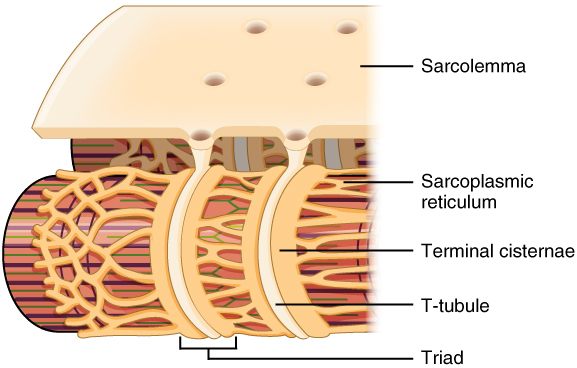
Figure 2. Narrow T-tubules permit the conduction of electrical impulses. The SR functions to regulate intracellular levels of calcium. Two terminal cisternae (where enlarged SR connects to the T-tubule) and one T-tubule comprise a triad—a "threesome" of membranes, with those of SR on two sides and the T-tubule sandwiched between them.
This process of sending a signal from the brain, across the neuromuscular junction, to cause contraction in the skeletal muscle is known as excitation-contraction coupling. And since we have discussed each section of this so far minus the actual steps of contraction (Sliding Filament Theory) let's summarize it with a series of steps that include the ones we have already mentioned earlier in the chapter.
Side note: creating this big picture in your head can often be the most difficult part of this chapter. I would encourage each of you to read each step and picture it visually in your head as you go through the steps. Do this several times a day for best success. This will help you get that big picture and understand this lengthy process.
Excitation – Contraction Coupling, in summary:
- Brain starts an action potential
- Action potential travels down the somatic motor neuron
- Action potential reaches the end of the neuron and opens voltage gated calcium channels on the synaptic end bulbs
- Calcium travels into the synaptic end bulb
- Calcium influx triggers ACh release out into the synaptic cleft by exocytosis
- ACh bind to a ligand-gated sodium channel on the motor end plate
- The ligand-gated sodium channel opens and sodium enters the skeletal muscle cell
- Sodium influx begins an action potential in the skeletal muscle cell
- The action potential travels along the sarcolemma
- The action potential travels down the T-tubules and activates the triad
- Calcium is released from the sarcoplasmic reticulum
- Calcium (the "key") binds to troponin (the "lock")
- Troponin moves and pulls tropomyosin off of the myosin binding sites "opening the door"
- With the binding sites now available the steps of the Sliding Filament Theory begin and contraction occurs.
![]()
![]()
Animation 6. Action Potential Travels the Sarcolemma. View the YouTube animation Muscle Excitation Part 5 (opens in new window).
Place the 5 given steps of excitation – contraction coupling in order or occurrence (not all steps are listed).
![]()

Continue your quiz here:

