
Layers of the Skin
This organ we call the skin (Figure 1) consists of two primary layers, the thinner, superficial epidermis and the thicker, deeper dermis (epi - upon or above; derma - skin). There is a third layer deep to the dermis called the hypodermis, but it is not technically part of the skin, also called the cutaneous membrane. The hypodermis is where we get the term hypodermic needle, which is obviously inserted " under " the dermis or skin. The hypodermis is also called the subcutaneous layer (you've heard of giving a shot " sub-Q ", this is where that term comes from- "subcu" or under the cutaneous membrane or skin).
(a)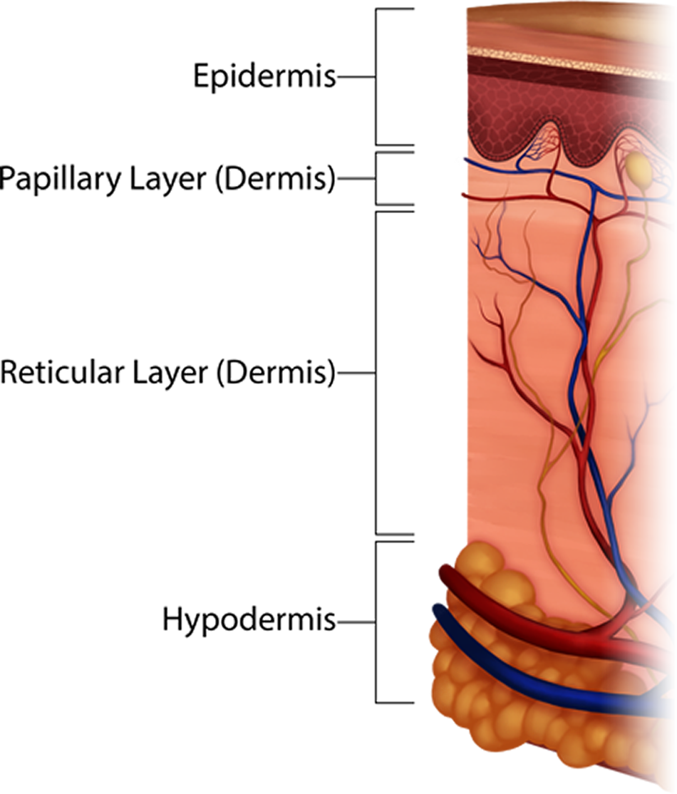 (b)
(b) 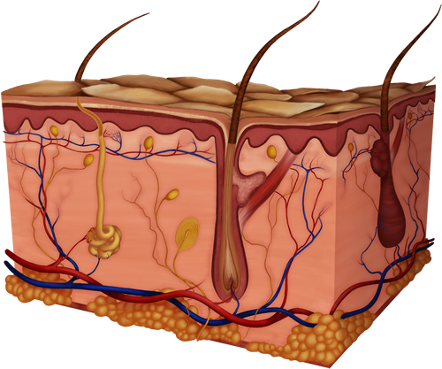
Figure 1. C ross-Sections illustrating skin layers: (a) is a smaller cross-section with the layers of skin (epidermis, the two layers of the dermis- papillary and reticular, and hypodermis) labeled; while (b) is a larger cross-section of skin and many of its internal structures. In this diagram you can tell that the epidermis is thin (shown in the darker colored layers at the top and only about 0.1mm deep!) and the dermis contains most of the skin's organs. The hypodermis is shown beneath the dermis, and is made of adipose tissue resembling groups of yellow balls.
Notice in the figure below that the surface layer, the epidermis, is not filled with blood vessels, or vascularized, at all. This serves a vital protective function for the body since the epidermis is commonly injured from even the simplest of daily activities. Imagine if the epidermis was vascularized, every paper cut could open a blood vessel and cause you to bleed profusely. The dermis, the deeper layer of skin, is well vascularized (has numerous blood vessels). This is important to not only receive nutrients but to place them in close enough proximity to allow nutrients to diffuse to the epidermis. Those cells stay alive even though it has no blood vessels of its own. Notice, also, the many nervous receptors in the skin, too, which keep communication lines open to the brain and provide information about our external environment.
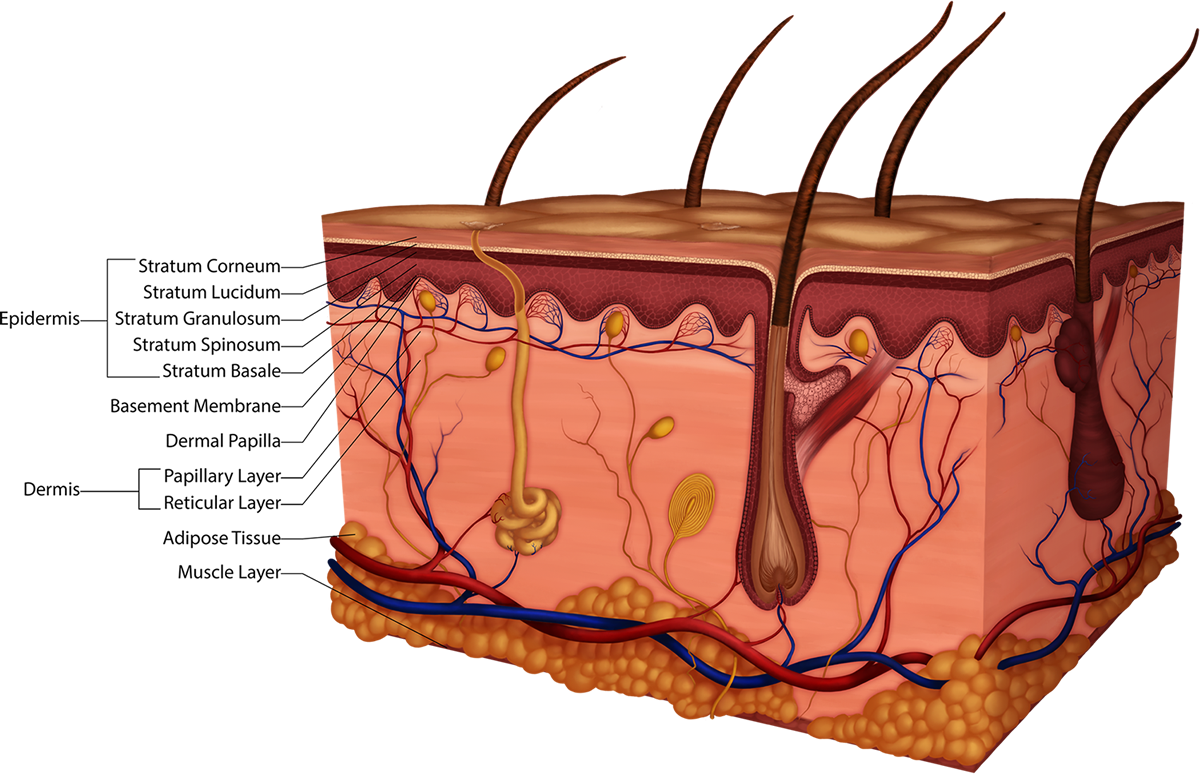
Figure 2. Parts of the Epidermis and the Dermis: Epidermal layers are the Stratum Corneum, Stratum Lucidum (only in thick skin), Stratum Granulosum, Stratum Spinosum and Stratum Basale (ba-sal-ee). The dermis has the superior papillary layer and the deeper reticular layer. Adipose is under these two layers of the skin and muscle is seen under that.
The epidermis is a stratified squamous epithelial tissue that has cells called keratinocytes, which are cells that eventually produce a very durable protein, keratin. Keratin is a water repellant protein that saturates the cells as they apporach the top layer, the corneum. Keratinization increases in the cells from deep to superficial, starting in the middle epidermal layer called stratum granulosum. The keratin protein is akin to the wax used on your car. Just like the wax, the protein repels water causing it to bead on the surface; however, your skin and the wax are not waterproof. If you soak in the tub initially water will bead on your skin, but eventually the epidermis will saturate and wrinkle or prune.
