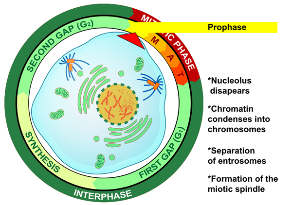
I. Prophase
At the beginning of prophase, the first stage of mitosis, the DNA condenses into chromosomes. Each chromosome holds two copies of each gene, with one gene on each chromatid. The two chromatids are also called sister chromatids. Sister chromatids are most closely connected at a region known as the centromere, which lies approximately in the center of the chromatids. When a chromosome condenses during prophase, it forms a shape similar to the one shown in the figure below. The two halves of chromosomes are sister chromatids, which are duplicate copies of one DNA strand. The two sister chromatids are held together by cohesions, which are proteins that form a temporary adhesive connection between the inner faces of the sister chromatids.

Figure 3. This illustration has a group of cells in prophase, one of which is shown with many blue chromosomes throughout the nucleus. The chromosome, enlarged and shown in blue, resembles the letter X. If you were to cut the chromosome in half through the centromere, each half is then referred to as a sister chromatid.
During prophase, the nuclear membrane, or nuclear envelope, disintegrates and the mitotic spindle assembles.

Figure 4. In this illustration of prophase, the first stage of mitosis, you can see that the nucleolus disappears, represented by the green dashed line around the nucleus. The chromatin also condenses into chromosomes, which is represented by the X shapes in the center in the color orange. Along the outer edge of the cell you can see the separation of entrosome and formation of the miotic spindle, shown in blue.