Objective
- Describe the neuronal components and functions of the posterior column-medial lemniscus pathway, the anterolateral pathway, and the spinocerebellar pathway.
Once any sensory cell transduces (converts) a stimulus into a nerve impulse, that impulse has to travel along sensory axons to reach the CNS. The nerves that convey sensory information from the periphery to the CNS are either spinal nerves, connected to the spinal cord, or cranial nerves, connected to the brain.
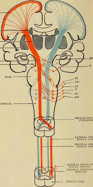
Generally, spinal nerves contain afferent axons from sensory receptors in the periphery, such as from the skin, mixed with efferent axons travelling to the muscles or other effector organs. As the spinal nerve nears the spinal cord, it splits into dorsal and ventral roots. The dorsal root contains only the axons of sensory neurons, whereas the ventral roots contain only the axons of the motor neurons. (Remember the words that go together section we already covered?) Typically, spinal nerve systems that connect to the brain are contralateral, in that the right side of the body is connected to the left side of the brain and the left side of the body to the right side of the brain. This crossing over of information from right body to left brain is termed decussation and can happen at two locations: the pyramids of the medulla oblongata and in the white matter of the spinal cord. In the simplest terms, the right hemisphere of the brain receives information from and controls motor output to the left side of the body, and vice versa for the left hemisphere of the brain. See the diagram below for a visual depiction of decussation.

Figure 4: Notice how the blue information originates and enters on the left side of the spinal cord and as it ascends, will decussate (cross over/switch side) at the pyramids of the medulla. This decussation means that information originating on the left side of the body is interpreted by the right side of the brain. The opposite is true is and illustrated by the orange lines. CCBY: Flickr API
Cranial nerves convey specific sensory information from the head and neck directly to the brain. Whereas spinal information is contralateral, cranial nerve systems are mostly ipsilateral, meaning that a cranial nerve on the right side of the head is connected to the right side of the brain. Some cranial nerves contain only sensory axons, such as the olfactory, optic, and vestibulocochlear nerves. Other cranial nerves contain both sensory and motor axons, including the trigeminal, facial, glossopharyngeal, and vagus nerves (however, the vagus nerve is not associated with the somatic nervous system). The general senses of somatosensation for the face travel through the trigeminal system.
.
A sensory pathway that carries peripheral sensations to the brain is referred to as an ascending pathway, or ascending tract. The various sensory modalities each follow specific pathways through the CNS. Somatosensory stimuli from below the neck pass along the sensory pathways of the spinal cord, whereas somatosensory stimuli from the head and neck travel through the cranial nerves. The somatosensory pathways are divided into two separate systems on the basis of the location of the receptor neurons. The dorsal column system (also referred to as the medial lemniscus pathway) and the spinothalamic tract are the two major pathways that bring sensory information to the brain. The sensory pathways in each of these systems are composed of three successive neurons.
Dorsal/Posterior Column System
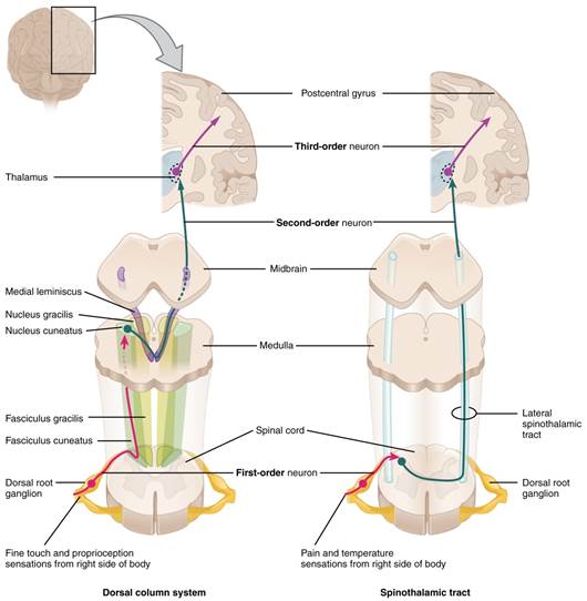
The dorsal column system begins with the axon of a dorsal root ganglion neuron entering the dorsal root and joining the dorsal column white matter in the spinal cord. This means that the dendrites of the first neuron in the dorsal column system receive the signal from the stimulated receptor. The action potential travels up the dendrites, into the spinal nerve, into the dorsal root, and then finally reaching the soma of the neuron in the dorsal root ganglion. Once the signal is processed by the soma of this unipolar neuron, the action potential travels down the axon from the dorsal root ganglion and into the posterior grey horn. The axon does not end here, but rather projects into the posterior white columns and ascends up the spinal cords into the medulla oblongata. Because this dorsal root ganglion neuron is the first to carry the information from receptor to higher processing in the cerebrum it is termed the first order neuron.
The next step in this system is for a multipolar neuron whose soma resides in the medulla, receives the information from the first order neuron and decussates – crosses the midline of the medulla - before continuing to ascend toward the cerebrum. Since this is the second neuron of the system, it is termed the second order neuron. Think of a relay race where one runner takes a lap around the track but then has to pass the baton to the next runner who will take their lap around the track. This is made possible because the first runner ends where the second runner begins, so the signal (the baton) can be passed efficiently from one runner to the next. This is very similar to how the somatosensory pathways operate. The first runner (first order neuron) brought the baton (signal) from the starting line (the receptor) into the medulla. This is where the second runner (second order neuron) picks up the baton and carries it toward the cerebrum to the next runner. These axons then continue to ascend the brain stem as a bundle called the medial lemniscus.
The second order neurons terminate in the thalamus, where each synapses with the third order neuron (third runner) in their respective pathway. The third neuron in the system projects its axons to the postcentral gyrus of the cerebral cortex also known as the primary sensory cortex, where somatosensory stimuli are initially processed and the conscious perception of the stimulus occurs.
Spinothalamic System
The spinothalamic tract begins with dorsal root ganglion neurons, the same type of first order neuron that was used and described in the dorsal column system. However, the axons of these first order neurons do not ascend the spinal cord, but rather they terminate in the posterior grey horn. This is where the baton is passed from the first order neuron to the awaiting second order neuron. The name "spinothalamic" comes from this second neuron, which has its cell body in the spinal cord gray matter and connects to the thalamus. Axons from these second order neurons then decussate within the spinal cord and ascend to the brain and enter the thalamus, where each synapses with the third neuron in its own pathway.
These two systems are similar in that they both begin with dorsal root ganglion cells, as with most general sensory information. The dorsal column system is primarily responsible for touch sensations and proprioception, whereas the spinothalamic tract pathway is primarily responsible for pain and temperature sensations. Another similarity is that the second neurons in both of these pathways are contralateral, because they project across the midline to the other side of the brain or spinal cord. In the dorsal column system, this decussation takes place in the brain stem; in the spinothalamic pathway, it takes place in the spinal cord at the same spinal cord level at which the information entered. The third neurons in the two pathways are essentially the same, synapsing in the thalamus, and the thalamic neuron projects to the somatosensory cortex. See the figure below for a side by side illustration of both systems.

Figure 5: The dorsal column system and spinothalamic tract are the major ascending pathways that connect the periphery with the brain. CCBY: OpenStax college
Diencephalon
The diencephalon is beneath the cerebrum and includes the thalamus and hypothalamus. In the somatic nervous system, the thalamus is an important relay for communication between the cerebrum and the rest of the nervous system. The hypothalamus has both somatic and autonomic functions. In addition, the hypothalamus communicates with the limbic system, which controls emotions and memory functions.
Sensory input to the thalamus comes from most of the special senses and ascending somatosensory tracts. The thalamus is a required transfer point for most sensory tracts that reach the cerebral cortex, where conscious sensory perception begins. The one exception to this rule is the olfactory system. The olfactory tract axons from the olfactory bulb project directly to the cerebral cortex. The thalamus is the phone operator of somatosensation. If you call a college campus but don't know the number for the bookstore, you will talk to the operator who will connect you to the right destination. The thalamus does this for sensory information; it takes the second order neuron, and says "How may I direct your call?" connecting it to the correct third order neuron so the information reaches the correct destination. The anterior thalamus serves as a relay between the hypothalamus and the emotion and memory-producing limbic system. The medial portion serves as a relay for information from the limbic system and basal ganglia to the cerebral cortex. This allows memory creation during learning, but also determines alertness. The special and somatic senses connect to the lateral portion, where their information is relayed to the appropriate sensory cortex of the cerebrum.
Cortical Processing
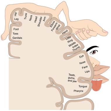
The cerebral cortex maintains the sensory topography of the body in reference to the position of the receptor cells. The somatosensory cortex (postcentral gyrus) provides an example in which, in essence, the locations of the somatosensory receptors in the body are mapped onto the somatosensory cortex. This mapping is often depicted using a sensory homunculus (see the figure below).
The term homunculus comes from the Latin word for "little man" and refers to a map of the human body that is laid across a portion of the cerebral cortex. In the somatosensory cortex, the external genitals, feet, and lower legs are represented on the medial face of the gyrus within the longitudinal fissure. As the gyrus curves out of the fissure and along the surface of the parietal lobe, the body map continues through the thighs, hips, trunk, shoulders, arms, and hands. The head and face are lateral to the fingers as the gyrus approaches the lateral sulcus. The representation of the body in this topographical map is medial to lateral from the lower to upper body. The connections through the thalamus maintain topography such that the anatomic information is preserved. Note that this correspondence does not result in a perfectly miniature scale version of the body, but rather exaggerates the more sensitive areas of the body, such as the fingers and lower face. Less sensitive areas of the body, such as the shoulders and back, are mapped to smaller areas on the cortex.

Figure 6: A cartoon representation of the sensory homunculus arranged adjacent to the cortical region in which the processing takes place. Note the exaggeration of the more sensitive body area ie. the size of the hand versus the elbow. CCBY: OpenStax college
Sensory input to the brain enters through pathways that travel through either the spinal cord (for somatosensory input from the body) or the brain stem (for everything else, except the visual and olfactory systems) to reach the diencephalon. In the diencephalon, sensory pathways reach the thalamus. This is necessary for all sensory systems to reach the cerebral cortex, except for the olfactory system that is directly connected to the frontal and temporal lobes.
The cortex has been described as having specific regions that are responsible for processing specific information; there is the visual cortex, somatosensory cortex, gustatory cortex, etc. However, our experience of these senses is not divided. Instead, we experience what can be referred to as a seamless percept. Our perceptions of the various sensory modalities—though distinct in their content—are integrated by the brain so that we experience the world as a continuous whole. In the cerebral cortex, sensory processing begins at the primary sensory cortex, then proceeds to an association area, and finally, into a multimodal integration area.


