

Anatomy of the Spinal Cord and Spinal Nerves
Objectives
By the end of this section, the student will be able to:
- Identify the major structures and regions of the spinal cord
- Identify the spinal nerves location, structure and function
- Explain the arrangement of gray and white matter in the spinal cord
- Explain the sensory and motor pathways of the spinal cord
- Understand important neural disorders
The Spinal Cord
The central nervous system (CNS) contains the brain and spinal cord, and the peripheral nervous system (PNS) is everything else. The spinal cord is the extension of nervous tissue in the vertebral column contained within the vertebral cavity. As the spinal cord continues to develop in the newborn, anatomical features mark its surface. For instance, the anterior midline is marked by the anterior median fissure, and the posterior midline is marked by the posterior median sulcus.
Spinal Cord Anatomy
The length of the spinal cord is divided into regions that correspond to the regions of the vertebral column. The name of a spinal cord region corresponds to the level at which spinal nerves pass through the intervertebral foramina. Immediately inferior to the brain stem is the cervical region, followed by the thoracic, then the lumbar, and finally the sacral region. The spinal cord is not the full length of the vertebral column because the spinal cord does not grow significantly longer after the first or second year, but the skeleton continues to grow. The nerves that emerge from the lower spinal cord pass through the intervertebral formina at the respective levels. As the vertebral column grows, these nerves grow with it and result in a long bundle of nerves that resembles a horse's tail and is named the cauda equina. The sacral spinal cord is at the level of the upper lumbar vertebral bones. The spinal nerves extend from their various levels to the proper level of the vertebral column.
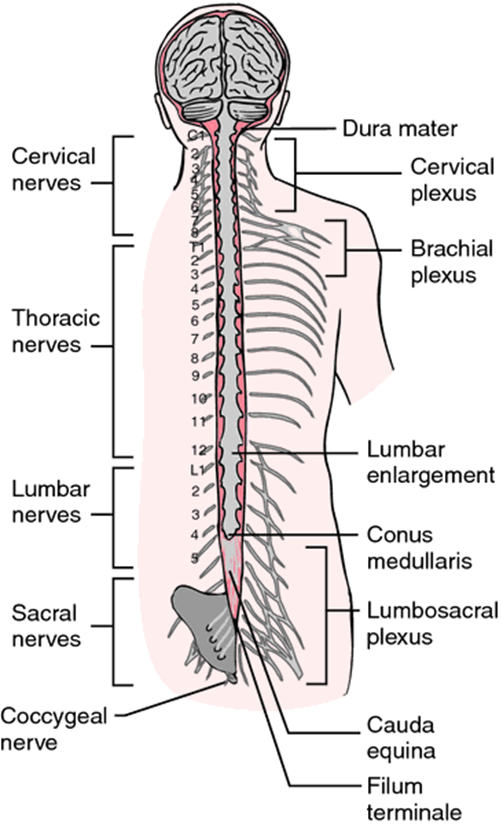
Figure 1. The spinal cord is not the full length of the vertebral column and it ends at the conus medullaris, usually around L1 or L2 on the vertebral column. The nerves that emerge from the lower spinal cord result in a long bundle of nerves that resembles a horse's tail and is named the cauda equina. The connective tissue anchor that extends from the conus medullaris down to the coccyx is the filum terminale, which helps to stabilize the cord in the subarachnoid space (along with denticulate ligaments that extend from the pia mater to the dura mater further anchoring the cord).
The end of the spinal cord containing cell bodies for motor neurons leaving the CNS is at the conus medullaris (about L1 to L2). This area is where the final nerves to and from the lower extremities enter or exit the spinal cord (remember that each spinal nerve contians both sensory and motor neurons communicating with the CNS). The next figure shows you the cauda equina of the spinal cord where the lumbar and sacral neurons extend from the end of the spinal cord and reach out (via intervertebral foramina) to the lower areas of the body.
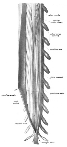
Figure 2. A dissection of the spinal cord showing the conus medullaris (the end of the spinal cord shaped like a cone in the middle of the nerves seen there), the cauda equina (looks like a horse's tail), and the filum terminale (the anchor to the spinal cord). By Dr. Johannes Sobotta - Sobotta's Textbook and Atlas of Human Anatomy 1908
