
What Does Simple Squamous Epithelium Do? What Does it Look Like?
Simple squamous epithelium is a very thin, flat-celled tissue. Because the cells are thin, this tissue is found where transport of chemical compounds needs to be fast. For example, this tissue is found in alveoli in your lungs, allowing oxygen to diffuse quickly.
The following video discusses simple squamous epithelium.
![]()
![]()
Video 3. View the Simple Squamous Epithelium video on youTube (opens in a new window).
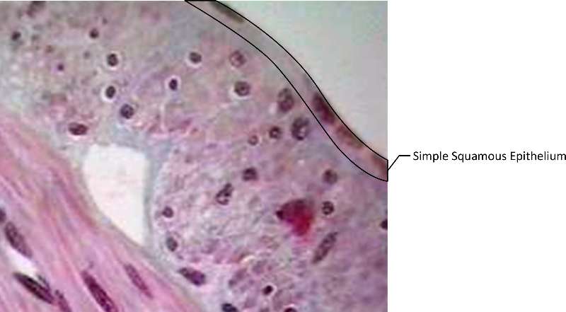
Figure 1. Cross section of small intestine (dyed purple for ease of visabilty). Simple Squamous Epithelium is outlined in black in the upper right corner.
Where is Simple Columnar Epithelium Found? What Does it Look Like?
The word simple tells you that simple columnar epithelium is composed of a single layer of cells. They are very tall and column shaped. Some of the cells are goblet cells, which aid in mucous secretion. Simple columnar epithelium is found lining the stomach, intestines and uterus. It functions to protect, secrete and absorb.
The following video discusses simple columnar epithelium.
![]()
![]()
Video 4. View the Simple Columnar Epithelium video on YouTube (opens in a new window)
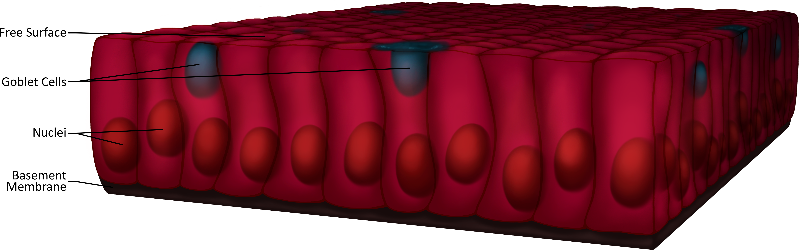
Figure 2. Cross section of simple columnar epithelium; The free surface, goblet cells, nuclei and basement membrane are labeled; note the column-shaped epithelial cells.
What is the Appearance and Role of Psuedostratified Columnar Epithelium with Cilia?
The prefix psuedo indicates that the tissue is not truly stratified. The varying locations of nuclei in this tissue gives the appearance of stratification, but the tissue is a single layer, simple tissue. Pseudostratified columnar epithelium with cilia also contains goblet cells, and as the name implies, has cilia. These aid in secretion and movement of cells and particles. This tissue is found in places that small particles or cells may need to be moved along, like the lining of the trachea and the fallopian tubes. The goblet cells secrete mucous which lubricates and protects the free surface.
The following video discusses psuedostratified columnar epithelium with cilia.
![]()
![]()
Video 5. View the Psuedostratified Columnar Epithelium video on YouTube (opens in a new window)
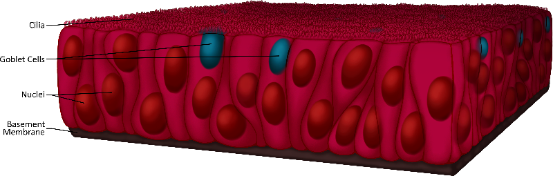
Figure 3. Cross section of pseudostratified columnar epithelium; The ciliated surface, goblet cells, nuclei and basement membrane are labeled; note the "semi-layering" of the column-shaped epithelial cells.
Where is Simple Cuboidal Epithelium Found? What Does it Look Like?
This tissue is made up of a single layer of cube shaped cells with their nuclei located in the middle. The functions include secretion and absorption and are found lining the ducts of glands, and kidney tubules.
The following video discusses simple cuboidal epithelium.
![]()
![]()
Video 6. View Simple Cuboidal Epithelium video on YouTube (opens in a new window).
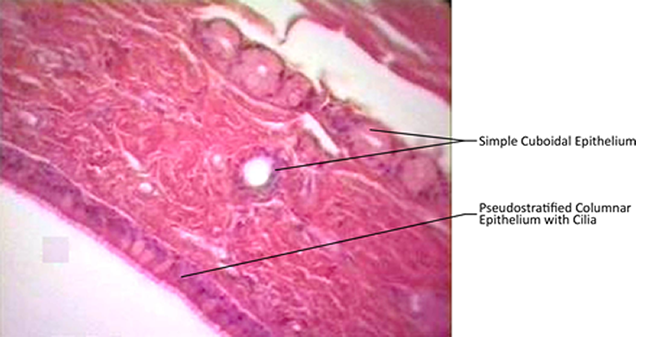
Figure 4. Micrographic trachea cross-section (dyed purple for ease of visabilty) with different types of epithelium labeled.
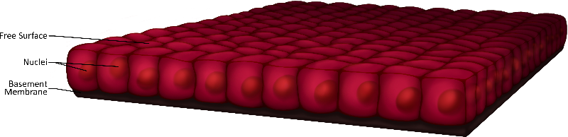
Figure 5. Simple cuboidal epithlium with free surface, nuclei and basement membrane labeled; note the cube-shaped cells.
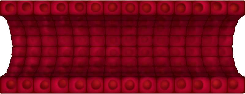
Figure 6. Longitudinal cross-section of a duct made of simple cuboidal epithelial tissue.
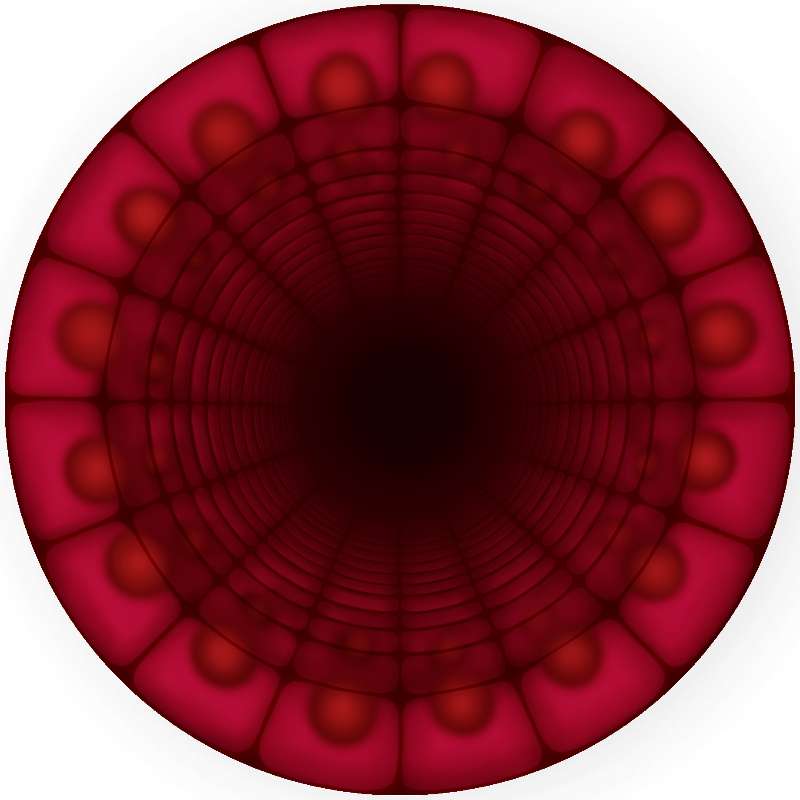
Figure 7. Cross-section of a duct made of simple cuboidal epithelial tissue.
What is Stratified Squamous Epithelial Tissue? What Does it Look Like and Where is it Found?
Stratified squamous epithelium has many layers of cells. The cells toward the free surface are flatter and those closer to the basement membrane are more cube-shaped. The main function of this tissue is protection. There are two kinds of stratified squamous epithelium: keratinized and nonkeratinized. The keratinized tissue is very tough because the cells contain the protein keratin. The cells in the outermost layer are dead, heavily keratinized cells. Keratininzed stratified squamous epithelium is found in your skin. Non keratinized stratified squamous epithelium is located in places such as the lining of the oral cavity and the rectum. This tissue is softer but its many layers of cells still serves to protect the underlying tissues. We will learn more about this tissue when we study the skin.
The following video discusses stratified squamous epithelial tissue.
![]()
![]()
Video 7. View the Stratified Squamous Epithelial Tissue video on YouTube (opens in a new window).
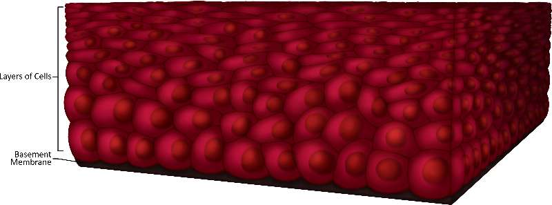
Figure 8. Cross-section of stratified squamous epitheial tissue; note the stacking of cells and the impact that has on their shape.
What is Transitional Epithelium? What Does it Look Like and Where is it Found?
Transitional epithelium appears to have few layers when it is stretched and many layers when it is not. It's found lining the bladder and when the bladder is full, that's when the tissue is stretched and appears to only have a few layers. When the bladder is emptied, the tissue appears to have many layers. Compare the two images below to see the difference.
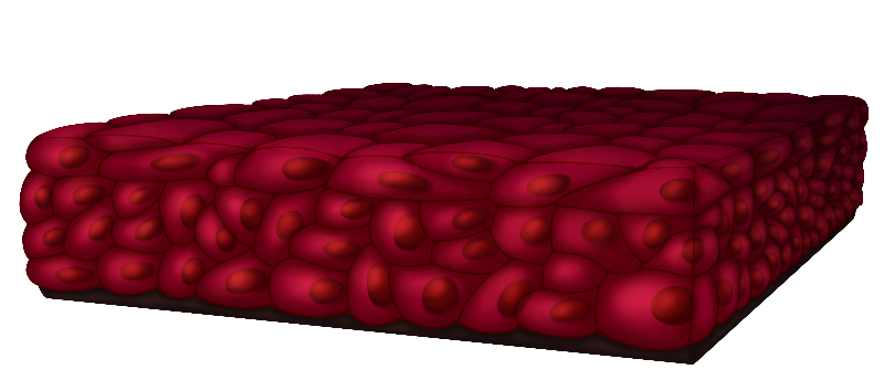
Figure 9. The image represents the changes that transitional epithelium tissues go through. Note the compactness of the cells.
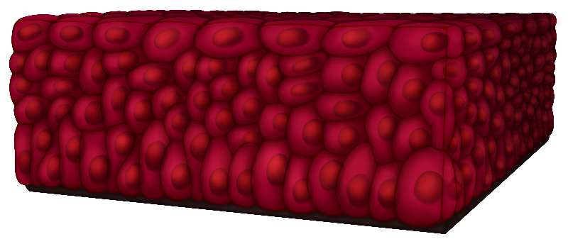
Figure 10. Represent the changes that transitional epithelium tissues go through. Note the the inflated-seeming cells.
The following video discusses transitional epithelium.
![]()
![]()
Video 8. View theTransitional Epithelium video on YouTube (opens in a new window).
Self-Check
![]()
| Drag the labels from the bottom to the correct slots. | |||
|
|||