
How are Organs Composed?
Organs are composed to two or more tissues working together to accomplish a particular function. Organs, such as membranes, can be as simple as an epithelial tissue and a connective tissue. For example, the conjunctiva that lines your eye lids are composed of simple squamous epithelium and loose connective tissue. Some organs are much more complicated. Consider the small intestine as an example. The small intestine is lined with simple columnar epithelium. The simple columnar epithelium protects the lining but it is also involved with absorption of the nutrients. Beneath the simple columnar epithelium is loose connective tissue. This tissue binds the layers together and is a good medium for the blood vessels that are there to absorb the nutrients and water that have been ingested. There are two layers of smooth muscle in the small intestine. The layer closest to the lumen encircles the lumen. When this layer contracts, the tube constricts. The next layer of smooth muscle runs the length of the intestine. When this longitudinal smooth muscle contracts, the tube shortens. The peristaltic action of the intestine occurs as these muscular layers contract. The small intestine is covered with a membrane composed of simple squamous epithelium and loose connective tissue. As you learn about the different organs in the human body, you will need to think about the tissues they are composed of and how the functions of the tissues allow the organs to perform their functions. Some examples of organs and their different tissues are labeled in the diagrams and micrographs below.
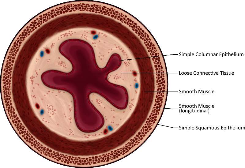
Figure 30. This is a very detailed illustration of the different tissue types fround in a cross-section of the small intestine. The burgundy starburst shape in the center is composed of simple columnar epithelium. The cream colored spongy structure outside of that is composed of loose connective tissue. The burgundy ring around that is composed of smooth muscle and the creme and burgundy very spongy ring outside of that is longitudinal smooth muscle. Lastly, the dark thin ring around the outside of the whole structure is composed of simple squamous epithelium.
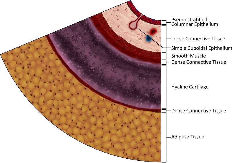
Figure 31. This image is a very detailed illustration of the different tissue types found in a cross-section of the trachea.The layer of the image, closest to this text, is yellow and globular adipose tissue and you can clearly see the fat cell membranes and shaded nuclei. The thick purple layer above that (which seems to contain bubbles) is hyaline cartilage, flanked on either side by thin layers of burgundy dense connective tissue. Above the top layer of dense connective tissue is a layer of burgundy smoth muscle. Atop that is a creme-colored spongy layer of loose connective tissue, intersected by a pink protrusion of simple cuboidal epithelium. Finally, all of these layers are topped by a thin layer of burgundy pseudostratified columnar epithelium.
