Introduction to the Appendicular Skeleton
- The student will be able to discuss the functions of the skeletal system.
- The student will be able to distinguish between the axial skeleton and appendicular skeleton.
- The student will be able to define the appendicular skeleton and its components.
- The student will be able to describe the bones and markings of the pectoral and pelvic girdles and how they connect limbs to the axial system.
- The student will be able to Identify the bones of the upper limbs and the major bony markings associated with each of the bones.
The appendicular skeletal system consists of the bones in the upper and lower appendages as well as those bones that connect them to the axial skeletal system. As you may recall from a previous lesson, the axial skeletal system consists of the main core of the body and includes the skull, thoracic (rib) cage, and vertebral column. The appendicular skeletal system can be subdivided into the pectoral girdle, the bones of the upper extremities, the pelvic girdle and the bones of the lower extremities.
When looking at the bones of the appendicular skeletal system keep in mind that there are two of each of the bones because there are appendages or limbs on both sides of the body. The right bones will be mirror images of the bones on the left. Also, it is helpful to look at the bones in all directions because there are distinct bone markings that you will find on the superior side, inferior side, anterior side, posterior side, lateral side, and medial side of each bone.
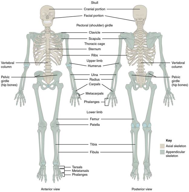
Figure 1: The axial skeleton supports the head, neck, back, and chest and thus forms the vertical axis of the body. It consists of the skull, vertebral column (including the sacrum and coccyx), and the thoracic cage, formed by the ribs and sternum, all of which touch the midline of the body or the axis. The appendicular skeleton is made up of all bones of the upper and lower limb, including the girdles that connect them to the axial skeleton.
The pectoral girdle consists of two bones, the scapula and clavicle. You can think of these two bones as being the connection between the axial skeleton and the upper arm, although only the clavicle directly connects with the axial skeletal system. The clavicle is located on the anterior side of the skeletal and has an S-shaped appearance to it and has two main connections. One of these connections is with the sternum of the axial system (called the sternal end) and the other connection is to the scapula (called the acromial end). The main purpose of the clavicle is as a support for the arms by holding the arms lateral to the body. If this support is damaged such as in a sports injury or car accident, then the muscles of the arm as well as the weight of the arm will be pulled forward, probably resulting in the person trying to hold the arm back. The most common fracture is in the thinner part of the shaft.
The scapula is what is commonly referred to as the shoulder blade. It is located posteriorly in the body and at a slight angle on the rib cage. It has two main skeletal interactions. One with the clavicle and one with the bone of the arm called the humerus. If looked at by itself, it has a shape of a triangle.
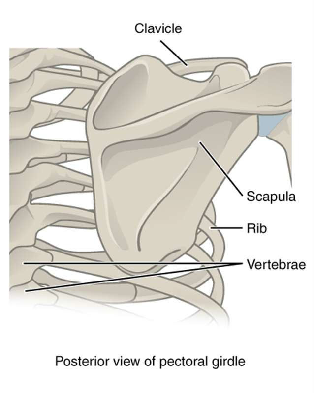
Figure 2: Posterior view of the Pectoral Girdle. The scapula, shaped like a triangle, is attached to the clavicle. The posterior side, seen here, shows the Scapular spine (the ridge across your shoulder blade) that ends superiorly at the acromion process (the point at the top of your shoulder).
On the anterior side superior side you can see the coracoid process (it has a "C" shape to it to help you remember the term has two c's), which is a point of attachment for a shoulder joint ligament. The supraspinous fossa, just posterior to the coracoid process, contains a muscle of a similar name and the subscapular fossa, which also contains a muscle, are seen from the anterior side. From the posterior side of the scapula you can see the acromion, which attaches to the clavicle and narrows at the spine. The spine of the scapula is the prominent ridge on the posterior side of the bone; the shallow depressions on either side of it are named appropriately. It separates the supraspinous fossa from an infraspinous fossa, which also contains a muscle. On the lateral side of the scapula is a shallow depression called the glenoid fossa or cavity, which contains the head of the humerus (the bone of the upper arm) and is part of the shoulder joint (glenohumeral joint). Overall the scapula is important for attachment of muscles especially for rotation at the shoulder that are collectively referred to as the rotator cuff and for connecting to the upper limb via the humerus. The acromioclavicular joint transmits forces from the upper limb to the clavicle. The ligaments around this joint are relatively weak. A hard fall onto the elbow or outstretched hand can stretch or tear the acromioclavicular ligaments, resulting in a moderate injury to the joint.
The following video discusses the pectoral girdle.
![]()
Video 1. View the Pectoral Girdle video on YouTube (opens in new window)