Pelvic Girdle

Figure 7. Bones of the Pelvic Girdle. [from openstax] The pelvic girdle is formed by the os coxae or hip bones. The hip bone attaches the lower limb to the axial skeleton through its articulation with the sacrum. The right and left hip bones, plus the sacrum and the coccyx, together form the pelvis.
The pelvic girdle consists of two bones called coxal bones. These bones serve as attachments for the lower limbs and to the axial skeleton through the sacrum. The two coxal bones are also attached to each other anteriorly. The bony pelvis is the entire structure formed by the two hip bones, the sacrum, and attached inferiorly to the sacrum, the coccyx. Unlike the bones of the pectoral girdle, which are highly mobile to enhance the range of upper limbe movements the bones of the pelvis are strongly united.
Unlike the bones of the pectoral girdle, which are highly mobile to enhance the range of upper limb movements, the bones of the pelvis are strongly united to each other to form a largely immobile, weight bearing structure. This is important for stability because it enables the weight of the body to be easily transferred laterally from the vertebral column, through the pelvic girdle and hip joints, and into either the lower limb lower limb whenever the other limb is not bearing weight. Thus, the immobility of the pelvis provides a strong foundation for the upper body as it rests on top of the mobile lower limbs.
The coxal bones are actually three bones that are fused into one structure. These three bones are called the ilium, ischium, and pubis. The superior part of the coxal bones is the flared part called the ilium. The ilium is connected to the sacrum in the posterior part of the hip called the sacroiliac joint. The inferior part of the coxal bone is composed of two bones, the ischium and the pubis. The ischium forms the posterior part of the inferior portion of the coxal bone. The pubis forms the anterior part of the inferior part of the coxal bone and is connected to each other at a joint called the pubic symphysis (symphysis pubis).
Ilium
The superior border of the ilium is the iliac crest that rounds to two iliac spines, which serve as attachment points for muscles. On the posterior margin is a U-shaped greater sciatic notch through which a nerve called the sciatic nerve passes.
Ischium
The ischium is the posterior inferior part of the coxal bone. The large, roughened area of the inferior ischium is the ischial tuberosity. This serves as the attachment for the posterior thigh muscles and also carries the weight of the body when sitting. You might think of this feature as the sitting bone. Another feature of the ischium is the ischial spine. This is a bony projection that separates two sciatic notches.
Pubis
This forms the anterior part of the inferior part of the pelvis. The pubic bones the two coxal bones are connected to each other through a joint called the pubic symphysis, which is slightly moveable but becomes more moveable during labor at the end of a pregnancy. When the pubic bones are connected to each other, you should also be able to detect an angle beneath the bones called the pubic arch or the pubic angle. The degree of the pubic arch differs depending on sex and is a useful indicator in forensics for determining whether a skeleton is male or female, as much of the difference between males and females is the structure of the pelvis as will be explained shortly.
Several features of the pelvis are formed from the fusion of all three bones or are best seen when the whole pelvis is put together. One of these features is a deep socket called the acetabulum. This is the articulation for the head of the femur and is considered one part of the hip joint. On the anteriorinferior is the obturator foramen. This opening contains connective tissue and muscles.
Spaces within the pelvis are divided into the greater pelvis (false pelvis) and the lesser pelvis (true pelvis). The greater pelvis encases some of the lower abdominal organs and extends from one ilium to the next ilium. The true pelvis is bordered by the pelvic brim and contains two openings. The opening into the true pelvis is called the pelvic inlet and the opening out of the true pelvis called the pelvic outlet.
|
Figure 8. Female Pelvis |
Figure 9. Male Pelvis |
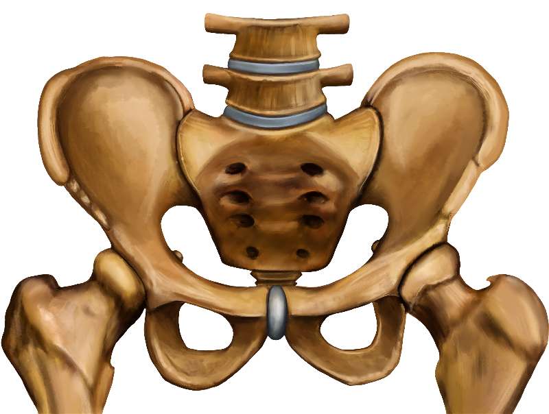
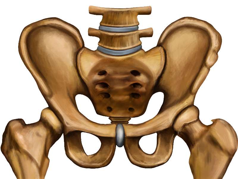
| Differences Between the Female and Male Pelvis | Female pelvis | Male pelvis |
|---|---|---|
| Pelvic weight | Bones of the pelvis are lighter and thinner | Bones of the pelvis are thicker and heavier |
| Pelvic inlet shape | Pelvic inlet has a round or oval shape | Pelvic inlet is heart-shaped |
| Lesser pelvic cavity shape | Lesser pelvic cavity is shorter and wider | Lesser pelvic cavity is longer and narrower |
| Pelvic arch | Pelvic arch is greater than 100 degrees | Pelvic arch is less than 90 degrees |
| Pelvic outlet shape | Pelvic outlet is rounded and larger | Pelvic outlet is smaller |
|
Greater Sciatic Notch |
Almost a 90 degree angle |
More narrow |
In general the major differences between the male and female pelvis, concern the female pelvis necessity of being wide enough for a baby to pass through. Therefore the female pelvis tends to be wider in both the pelvic inlet and pelvic outlet and tends to have a wide pubic arch of a 100 degrees or more where the male pelvis tends to be thicker because of the generally more muscular aspect of the male and has a pubic arch of less than 90 degrees.