The bones of the lower limbs include the femur, the tibia, the tarsals, metatarsals, and phalanges. Except for the tarsals, all of the bones of the lower limbs consist of long bones that have two distinct ends (epiphyses) and a shaft region called a diaphysis.
Femur
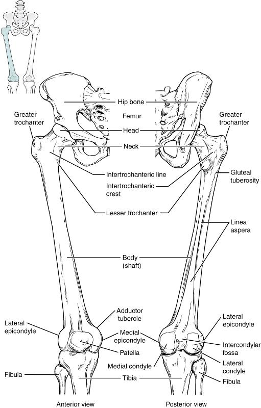
Figure 10. The bone of the thigh is called the femur. This is the longest bone and strongest bone of the body because of its need to hold up the entire weight of the body. This bone articulates with the acetabulum of the pelvis at its proximal end and the tibia at its distal end. It is often a site of many hip fractures. Several parts of the femur include the ball-shaped head, which articulates with the acetabulum and also include a small depression called the fovea capitis, which contains a ligament that helps to attach it to the acetabulum. Laterally next to the head are two large projections: the greater trochanters and lesser trochanters. These projections are attachments points for muscles or ligaments. The head is attached to the shaft of the bone by a neck. Along the shaft on the posterior side is the linea aspera, that is a site of attachment for the gluteal muscles. At the distal end of the femur are two condyles that articulate with the major bone of the lower leg called the tibia. Next to each of these condyles are epicondyles that provide attachment points for ligaments and tendons. Also articulating with the femur is the patella, which is a round-shaped bone that develops after birth and helps to provide leverage to the knee joint eventually helping the individual to walk.
Patella
The patella is a round (sesamoid) bone that is located inside of tendon. This tendon extends from the quadriceps muscle and attaches to the tibial tuberosity on the tibia. The patella helps to prevent this tendon from rubbing against the surface of the femur and also increases leverage during walking.
Tibia
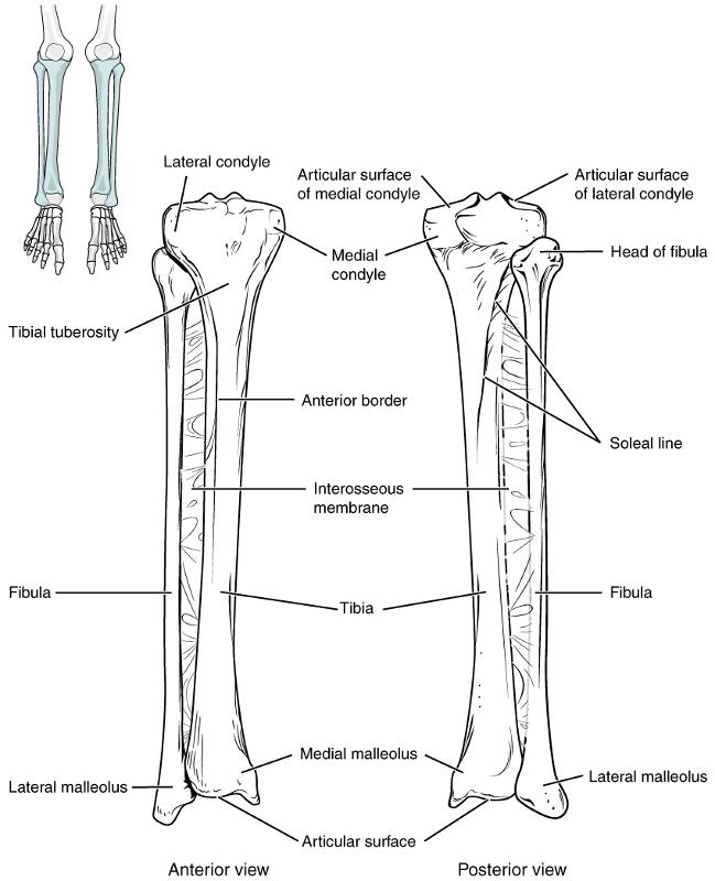
Figure 11. This is the medial bone of the lower leg. Of the two bones in the lower leg, the tibia bears most of the weight coming from the femur. At the proximal end of the tibia are two condyles that also articulate with the condyles of the femur. Below the condyles is a rough process called the tibial tuberosity. This process is a site of attachment for a tendon that connects the tibia with the patella and is a major ligament associated with the knee joint. Along the anterior side of the tibia is the anterior crest, which can be felt underneath the skin as a ridge. At the distal side on the medial side is the medial malleolus, which is large bony bump that is often thought of as the inner ankle.
Fibula
This is the lateral bone of the lower leg. In comparison with the tibia, the fibula is thinner and is not suitable for weight bearing. It is mostly for the attachment of various muscles and ligaments. At the proximal end of the fibula is the head of the fibula. his is a knobby structure that articulates with the proximal, lateral end of the tibia at the lateral condyle of the tibia. At the opposite end of the fibula is the lateral malleolus. This is a process that is easily felt on the lateral side of the ankle region. Along with the medial malleolus of the tibia, the lateral malleolus articulates with a large tarsal bone called the talus, which anchors the lower leg with the ankle and foot.