Humerus
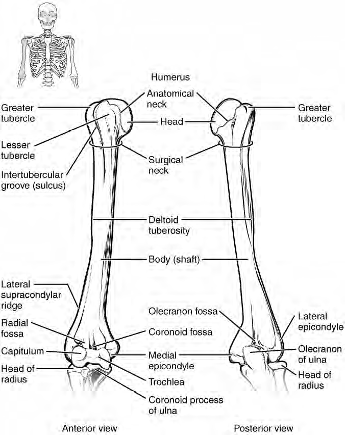
Figure 3. Humerus or upper arm bone. Both the right and left humerus bones are shown. Notice the proximal end's head of the humerus, the surgical neck (easier to cut or break than the anatomical neck), the greater tubercle on the lateral side, the deltoid tuberosity, the olecranon fossa, the lateral and medial epicondyles and the distal condyle (has two parts: trochlea and capitulum).
There is one bone in the arm region called the humerus. The humerus is a long bone that has two distinct bulbous ends called the epiphyses and a middle region generally called the diaphysis. At the proximal epiphysis, is a rounded ball shaped area called the head of the humerus. This feature fits in with the glenoid cavity of the scapula and is one part of what is called the shoulder joint. Next to the head of the humerus is the anatomical neck. Located on the lateral side of the proximal epiphysis is an expanded area called the greater tubercle and a smaller lesser tubercle that are separated from each other by the intertubercular groove. Both the greater tubercle and lesser tubercle are attachment points for muscles that act across the shoulder joint. The surgical neck is located at the base of the proximal epiphysis and attaches to the diaphysis. The surgical neck is commonly a site of arm fractures. On the diaphysis, is a roughed area called the deltoid tuberosity, which is a site of attachment for a muscle called the deltoid. On the distal epiphysis of the humerus is located a condyle. This condyle is divided up into two parts. The medial part of the condyle is called the trochlea, this is a pulley-shaped region which articulates with the trochlear notch of the ulna. The lateral part of the condyle is the capitulum, a rounded, knob-like structure that articulates with the radius bone. On the posterior distal epiphysis, is a deep depression called olecranon fossa that receives the olecranon process of the ulna and allows movement at the elbow joint as the arm extends.