Ulna and Radius
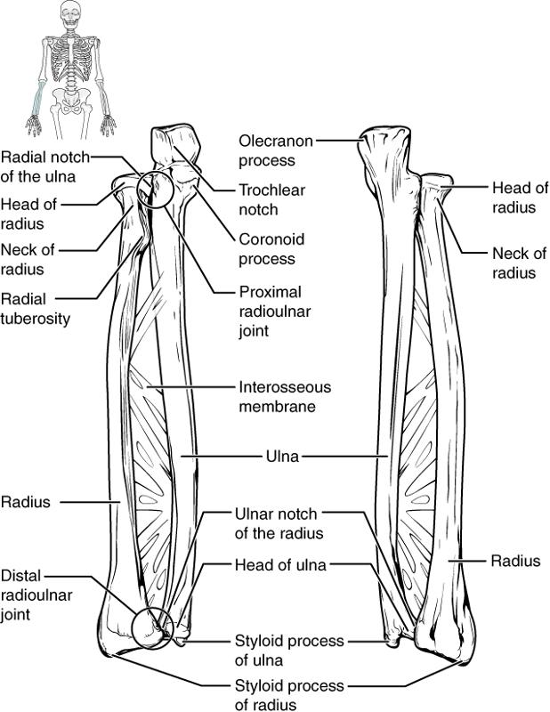
Figure 4. Ulna and radius of the lower arm.
The ulna is located on the medial side of the forearm, and the radius is on the lateral side. These bones are attached to each other by an interosseous membrane.On the ulna several landmarks (bone markings) are noticeable. The olecranon process that fits into the fossa on the humerus is our elbow. The trochlea of the humerus fits into the trochlear notch on the proximal side of the ulna, as well. The head of the radius is lateral to the ulna's coronoid process. On the radius make note of the radial tuberosity, since the biceps tendon inserts at this point (seen later in the muscles).
Carpals, Metacarpals, and Phalanx Bones (plural phalanges)
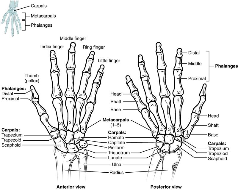
Figure 5. The wrist consists of eight carpal bones that are arranged in two rows. The carpal bones form the base of the hand. Three of these carpal bones articulate with radius. The ulna does not directly articulate with any of the carpal bones. The distal carpal bones articulate with each other and along with the carpals that articulate with the radius contribute to the movement of the hand at the wrist. The distal carpal bones also articulate with the metacarpal bones of the hand.
The main part of the hand consists of 5 metacarpal bones. These bones connect with the carpal bones as well as with phalanges of the fingers and thumbs. The proximal end of the each metacarpal bone articulates with one of the distal carpal bones. Each of these articulations is a carpometacarpal joint. The expanded distal end of each metacarpal bone articulates at the metacarpophalangeal joint with the proximal phalanx bone of the thumb or one of the fingers. The distal end also forms the knuckles of the hand, at the base of the fingers. The metacarpal bones are numbered 1-5 beginning at the thumb. The first metacarpal has greater flexibility than the other metacarpals, which allows us to grip objects with opposable thumbs.
The fingers and thumbs are made up of 14 bones each of which are called a phalanx. The plural form of phalanx is called phalanges. The phalanges are identified by their location. Each finger and thumb contain a proximal phalanx that articulates with a metacarpal bone. Each finger but not the thumb contains a middle phalanx. The fingers and thumb also contain distal phalanges. Therefore the fingers contain three phalanges each and the thumb contains two phalanges each.

Figure 6. In the articulated hand, the carpal bones form a U-shaped grouping. A strong ligament called the flexor retinaculum spans the top of this U-shaped area to maintain this grouping of the carpal bones. Together the carpal bones and the flexor retinaculum form a passageway called the carpal tunnel, with the carpal bones forming the walls and floor, and the flexor retinaculum forming the roof of this space. The tendons of nine muscles of the anterior forearm and a nerve pass through this narrow tunnel to enter the hand.
The carpal bones form a U-shape and a strong ligament called the flexor retinaculum spans the top of this U-shaped area to secure the carpal bones. Together the carpal bones and the flexor retinaculum form a passageway called the carpal tunnel. Overuse of the muscle tendons or wrist injury can produce inflammation and swelling within this space. This produces compression of the nerve, resulting in carpal tunnel syndrome, which is characterized by pain or numbness, and muscle weakness in those areas of the hand supplied by this nerve.